File list
This special page shows all uploaded files.
| Date | Name | Thumbnail | Size | Description | Versions |
|---|---|---|---|---|---|
| 17:19, 8 August 2013 | Test.mp4 (file) | 4.17 MB | More testing! | 2 | |
| 14:52, 8 August 2013 | Test.mp3 (file) | 5.65 MB | Testing out Html5mediator | 1 | |
| 03:43, 21 August 2013 | IPLab9RMSF8.jpg (file) |  |
49 KB | This is a high-power photomicrograph of a thrombosed vessel in the dermis. Note that the endothelial cells are missing along part of the circumference of the vessel (arrows)--this is where the main part of the thrombus has attached. Also note the infla... | 1 |
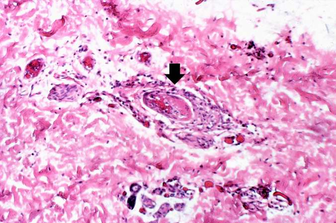
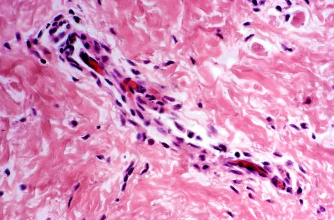
| 03:43, 21 August 2013 | IPLab9RMSF7.jpg (file) |  |
60 KB | This is a high-power photomicrograph of a dermal vessel (arrow) which is exhibiting vasculitis and thrombosis. | 1 |
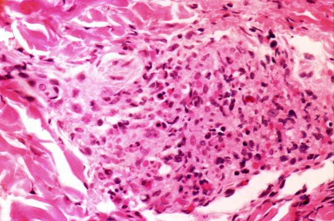
| 03:43, 21 August 2013 | IPLab9RMSF6.jpg (file) |  |
57 KB | This is a high-power photomicrograph of severe vasculitis in the dermis. | 1 |
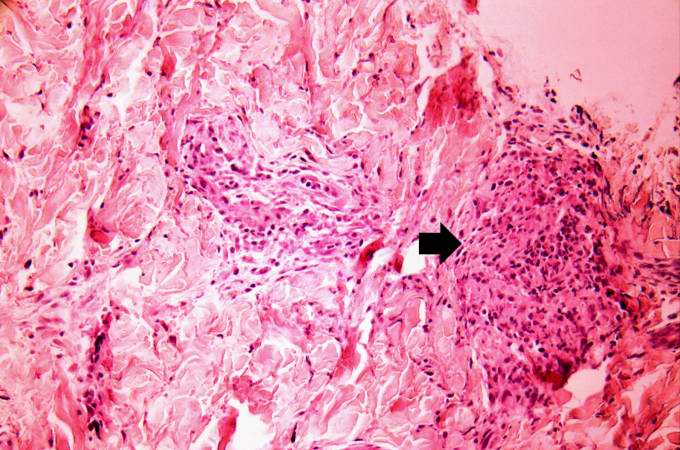
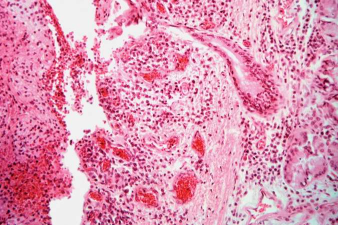
| 03:42, 21 August 2013 | IPLab9RMSF5.jpg (file) |  |
70 KB | This is a photomicrograph of dermis with an area of more severe vasculitis (arrow). | 1 |
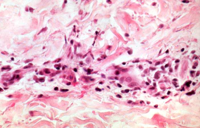
| 03:42, 21 August 2013 | IPLab9RMSF4.jpg (file) |  |
47 KB | This is a higher-power photomicrograph of a dermal vessel with mild vasculitis. | 1 |
| 03:42, 21 August 2013 | IPLab9RMSF3.jpg (file) |  |
52 KB | This is a photomicrograph of a small vessel in the dermis which is demonstrating mild vasculitis. | 1 |
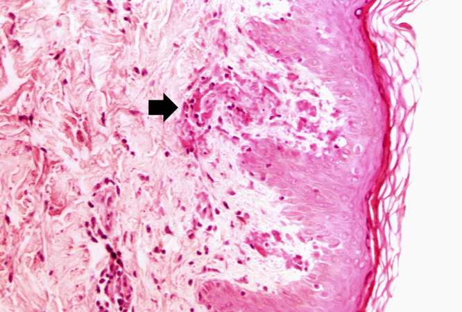
| 03:42, 21 August 2013 | IPLab9RMSF2.jpg (file) |  |
51 KB | This is a higher-power photomicrograph demonstrating areas of hemorrhage immediately underneath the epidermis. Also note the cellularity and thrombosis of the small vessels in the dermis (arrow). | 1 |
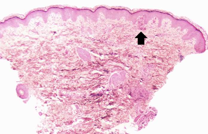
| 03:42, 21 August 2013 | IPLab9RMSF1.jpg (file) |  |
46 KB | This is a low-power photomicrograph of the skin biopsy. Note areas of hemorrhage (arrow) in the dermis. | 1 |
| 03:50, 21 August 2013 | IPLab9Diphtheria4.jpg (file) |  |
69 KB | In this higher-power photomicrograph of the tissue from the previous image, the ulcerated tracheal mucosa and the diphtheritic membrane are more clearly seen. Although difficult to make out at this magnification, most of the cells in this inflammatory ... | 1 |
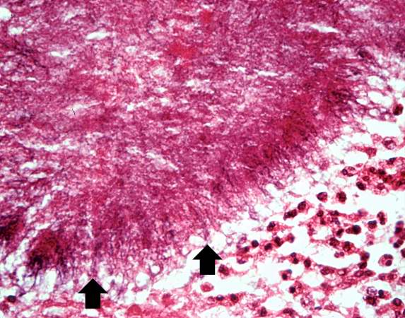
| 03:50, 21 August 2013 | IPLab9Diphtheria3.jpg (file) |  |
71 KB | This is an even higher-power photomicrograph of the tracheal mucosa and the diphtheritic membrane. The mucosal surface of the trachea is ulcerated (total loss of epithelial cells) and the only remaining epithelial cells are found in the glands (arrows)... | 1 |
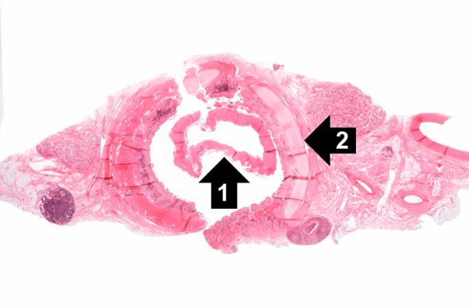
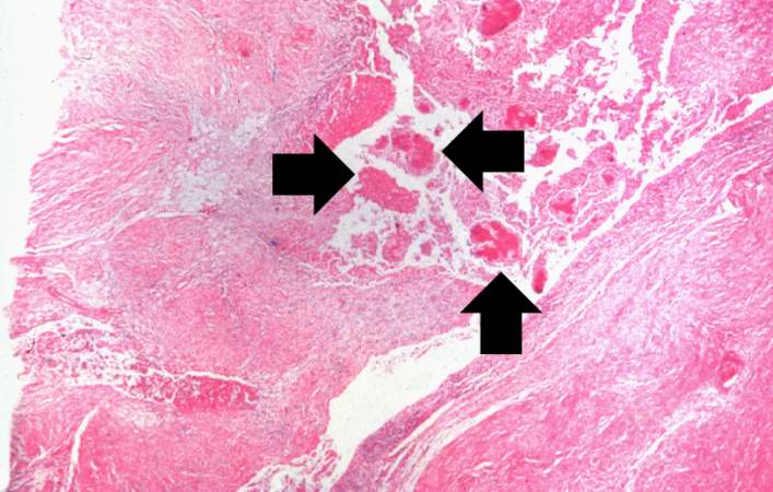
| 03:49, 21 August 2013 | IPLab9Diphtheria2.jpg (file) |  |
47 KB | This is a higher-power photomicrograph of trachea with the diphtheritic membrane (1). Even though, the main part of the membrane has pulled away from the tracheal lining during histological processing, in this section part of the membrane is still loos... | 1 |
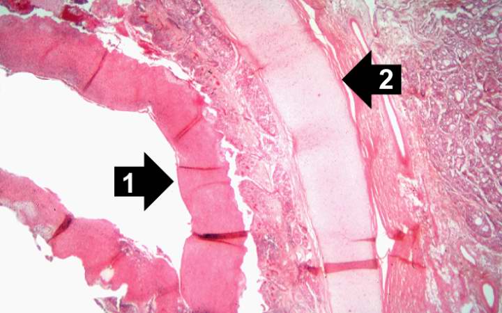
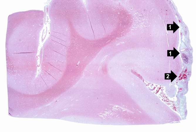
| 03:49, 21 August 2013 | IPLab9Diphtheria1.jpg (file) |  |
28 KB | This is a low-power photomicrograph of the trachea with the diphtheritic membrane (1), which has pulled away from the tracheal lining during histological processing. Note the tracheal cartilage (2) present in this section. | 1 |
| 03:57, 21 August 2013 | IPLab9Clostridium7.jpg (file) |  |
70 KB | This is a high-power photomicrograph of a tissue section stained with a tissue Gram's stain (Brown & Brenn). The Gram-positive bacilli can be seen throughout this tissue section. | 1 |
| 03:57, 21 August 2013 | IPLab9Clostridium6.jpg (file) |  |
55 KB | This higher-power photomicrograph of the previous image provides a clearer view of gas bubbles in the tissue, the necrotic hypereosinophilic muscle cell (1), and the mild inflammatory reaction (2). At this magnification, the bacteria located throughout... | 1 |
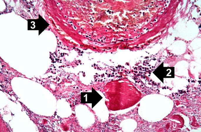
| 03:56, 21 August 2013 | IPLab9Clostridium5.jpg (file) |  |
68 KB | This high-power photomicrograph shows the gas accumulation present in the tissue, a necrotic muscle cell (1), and a mild inflammatory response (2). There is also a thrombosed blood vessel (3). The blue-staining rods (bacterial organisms) can barely be ... | 1 |
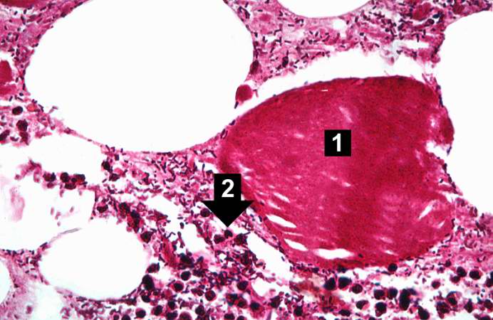
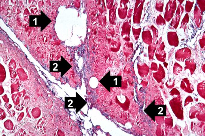
| 03:56, 21 August 2013 | IPLab9Clostridium4.jpg (file) |  |
83 KB | This is a high-power photomicrograph of skeletal muscle. The muscle cells are hypereosinophilic and most do not contain nuclei, indicating that these cells are dead or dying. The round clear spaces (1) in this tissue correspond to gas accumulations pri... | 1 |
| 03:56, 21 August 2013 | IPLab9Clostridium3.jpg (file) |  |
62 KB | This is a low-power photomicrograph of muscle fascicles containing large gas bubbles (arrows). Note that there is no inflammatory reaction in this section. | 1 |
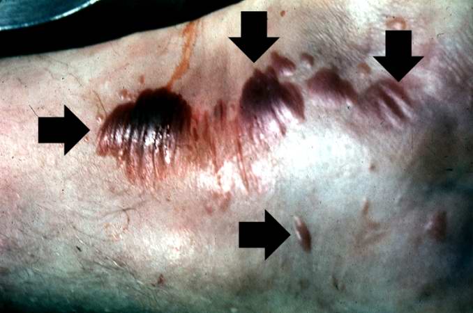
| 03:56, 21 August 2013 | IPLab9Clostridium2.jpg (file) |  |
40 KB | This gross photograph shows a close-up view of hemorrhagic blebs (arrows) on the skin. The blebs on the skin are accumulations of gas being discharged into the tissues from the Clostridium perfringens. This gas produces crepitance. | 1 |
| 03:56, 21 August 2013 | IPLab9Clostridium1.jpg (file) |  |
36 KB | This gross photograph of the lower extremity was taken at autopsy. Notice the swelling and the area of the primary infection (arrow). | 1 |
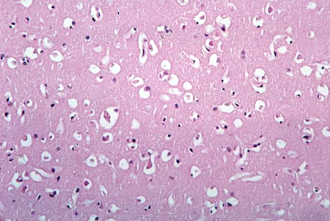
| 03:47, 21 August 2013 | IPLab9BacterialMeningitis8.jpg (file) |  |
56 KB | This photomicrograph of brain tissue demonstrates diffuse edema. | 1 |
| 03:47, 21 August 2013 | IPLab9BacterialMeningitis7.jpg (file) |  |
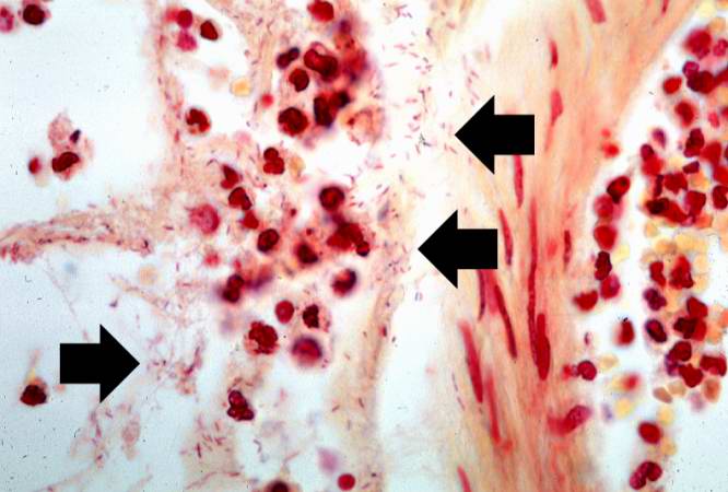
42 KB | This is a high-power photomicrograph of exudate from the leptomeninges which has been Gram-stained. Note the Gram-negative bacteria (arrows) throughout this section. | 1 |
| 03:47, 21 August 2013 | IPLab9BacterialMeningitis6.jpg (file) |  |
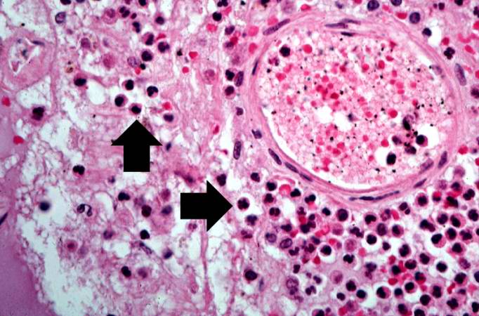
63 KB | This is a high-power photomicrograph of a blood vessel from the previous image. The vessel is surrounded by neutrophils (arrows). | 1 |
| 03:47, 21 August 2013 | IPLab9BacterialMeningitis5.jpg (file) |  |
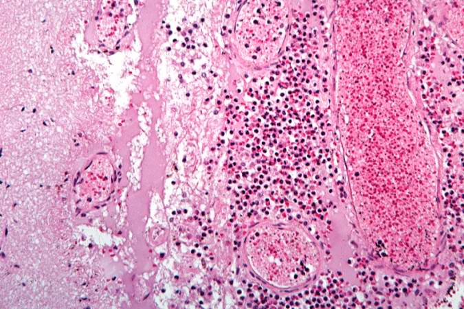
78 KB | This is a higher-power photomicrograph of inflammatory exudate in a sulcus. The majority of cells in this exudate are neutrophils. There is also abundant fibrin (arrows) and red blood cells are present in the congested vessels. | 1 |
| 03:46, 21 August 2013 | IPLab9BacterialMeningitis4.jpg (file) |  |
52 KB | This higher-power photomicrograph of a sulcus shows the congested vessels and the inflammatory exudate in the leptomeninges. | 1 |
| 03:46, 21 August 2013 | IPLab9BacterialMeningitis3.jpg (file) |  |
79 KB | This is a higher-power view of a congested blood vessel. Inflammatory exudate is present within the vessel and throughout the leptomeninges. | 1 |
| 03:46, 21 August 2013 | IPLab9BacterialMeningitis2.jpg (file) |  |
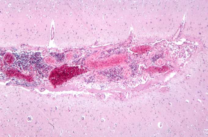
30 KB | This is a low-power photomicrograph of brain section. Note the exudate (1) in the meninges and congestion of the vessels (2) in the leptomeninges. | 1 |
| 03:46, 21 August 2013 | IPLab9BacterialMeningitis1.jpg (file) |  |
44 KB | This gross photograph of the autopsy specimen from this case demonstrates the purulent exudate (arrows) in the leptomeninges. | 1 |
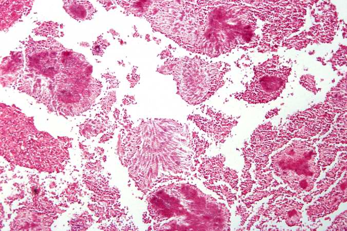
| 03:59, 21 August 2013 | IPLab9Actinomycosis5.jpg (file) |  |
63 KB | This is a high-power photomicrograph of an actinomycotic colony. The filamentous nature (arrows) of the actinomyces organisms is more easily appreciated at this power. | 1 |
| 03:59, 21 August 2013 | IPLab9Actinomycosis4.jpg (file) |  |
63 KB | This is an even higher-power photomicrograph of actinomycotic colonies in the abscess. The filamentous nature (arrows) of the actinomyces organisms in these colonies can be appreciated. | 1 |
| 03:59, 21 August 2013 | IPLab9Actinomycosis3.jpg (file) |  |
86 KB | This is a higher-power photomicrograph of actinomycotic colonies in the abscess. | 1 |
| 03:59, 21 August 2013 | IPLab9Actinomycosis2.jpg (file) |  |
59 KB | This is a higher-power photomicrograph of an abscess demonstrating a pocket of purulent exudate that contains numerous actinomycotic colonies (arrows). | 1 |
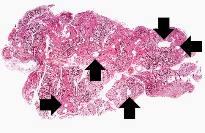
| 03:59, 21 August 2013 | IPLab9Actinomycosis1.jpg (file) |  |
39 KB | This is a low-power photomicrograph of the retroperitoneal abscess. At this magnification, multiple dark-staining foci can be appreciated. These foci are Actinomyces colonies (arrows). These colonies are known as "sulfur granules" because in gross spec... | 1 |
| 03:53, 21 August 2013 | IPLab9ARF6.jpg (file) |  |
50 KB | This high-power photomicrograph of myocardium shows the cellular detail of another Aschoff body. In this case there appears to be a multinucleated Aschoff giant cell (arrow). | 1 |
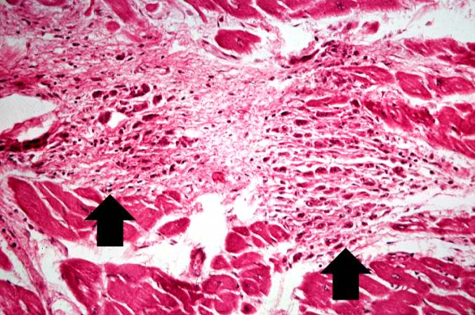
| 03:52, 21 August 2013 | IPLab9ARF5.jpg (file) |  |
52 KB | This high-power photomicrograph of myocardium shows the cellular detail of an Aschoff body. Aschoff bodies are foci of fibrinoid necrosis surrounded by lymphocytes, macrophages, an occasional plasma cell, and plump “activated” histiocytes called An... | 1 |
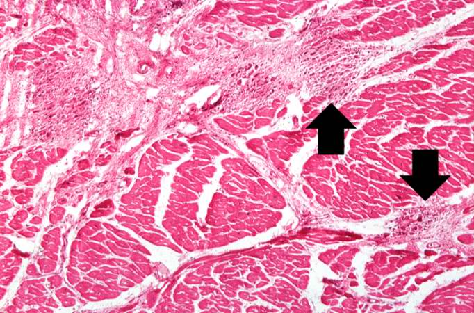
| 03:52, 21 August 2013 | IPLab9ARF4.jpg (file) |  |
76 KB | This is a higher-power photomicrograph of myocardium containing Aschoff bodies (arrows) within the interstitium. | 1 |
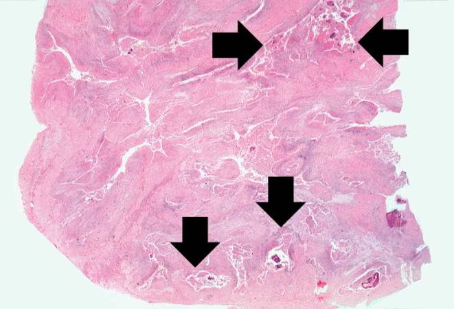
| 03:52, 21 August 2013 | IPLab9ARF3.jpg (file) |  |
83 KB | This is a higher-power photomicrograph of myocardium showing cellular accumulations--Aschoff bodies (arrows)--within the interstitium of the myocardium. These are found especially around blood vessels. | 1 |
| 03:52, 21 August 2013 | IPLab9ARF2.jpg (file) |  |
29 KB | This is a low-power photomicrograph of heart tissue. Little can be seen at this magnification, except that the tissue looks relatively normal. | 1 |
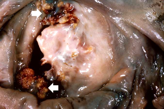
| 03:52, 21 August 2013 | IPLab9ARF1.jpg (file) |  |
48 KB | This is a gross photograph of mitral valve demonstrating marked thickening and fibrosis of the valve leaflet. There are also numerous foci of fibrinoid necrosis within the cusps and friable vegetations (verrucae) along the lines of closure (arrows). Th... | 1 |
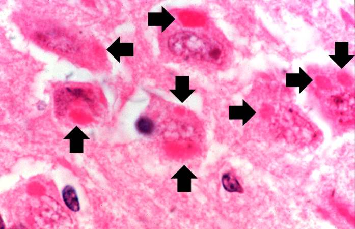
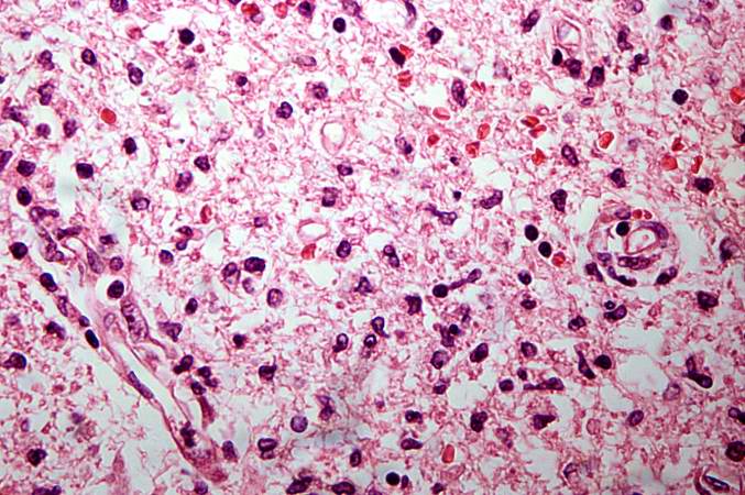
| 02:41, 21 August 2013 | IPLab8Rabies5.jpg (file) |  |
41 KB | This is a high-power photomicrograph of a neuron surrounded by inflammatory cells (lymphocytes and microglia). This neuron has two intracytoplasmic eosinophilic inclusion bodies (arrows). | 1 |
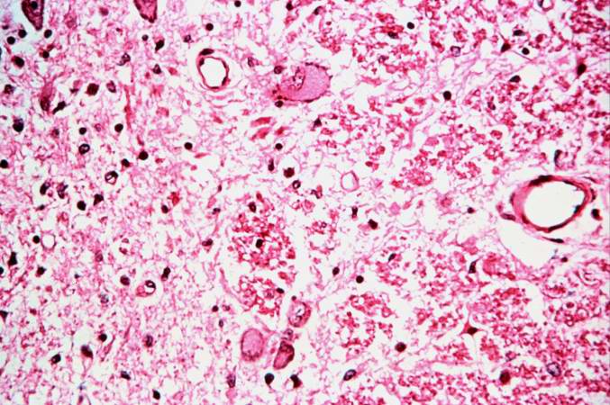
| 02:40, 21 August 2013 | IPLab8Rabies4.jpg (file) |  |
44 KB | This is a high-power photomicrograph of neurons containing variably-sized intracytoplasmic eosinophilic inclusion bodies (arrows). | 1 |
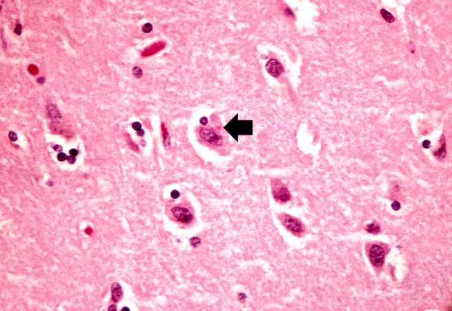
| 02:40, 21 August 2013 | IPLab8Rabies3.jpg (file) |  |
49 KB | This is a higher-power photomicrograph of the shrunken neurons. One neuron appears to have an eosinophilic intracytoplasmic inclusion body (arrow). | 1 |
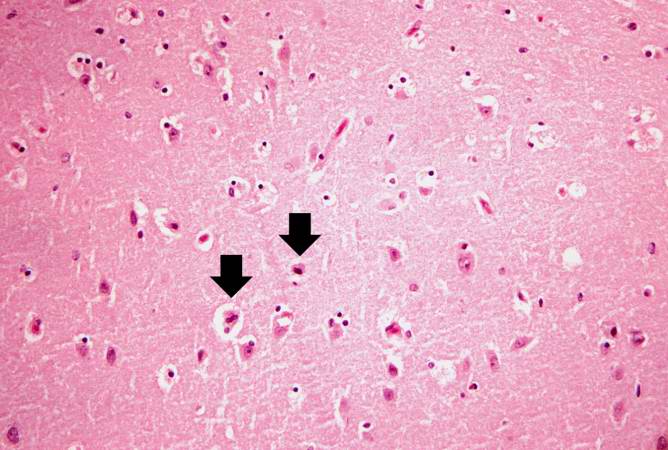
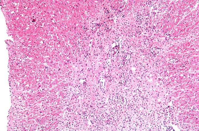
| 02:40, 21 August 2013 | IPLab8Rabies2.jpg (file) |  |
52 KB | This is a medium-power photomicrograph of brain tissue exhibiting edema and evidence of shrunken, necrotic neurons (arrows). | 1 |
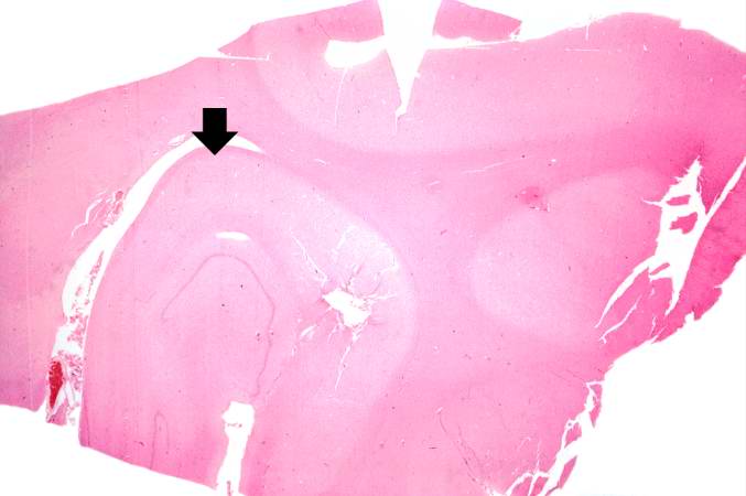
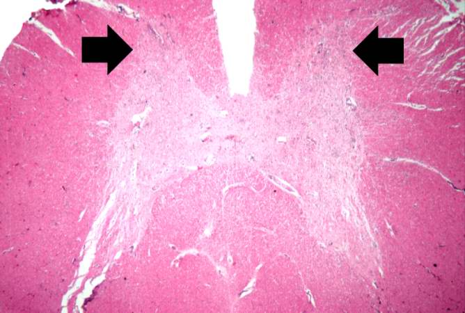
| 02:39, 21 August 2013 | IPLab8Rabies1.jpg (file) |  |
25 KB | This is a low-power photomicrograph of the hippocampus (arrow) from this case. | 1 |
| 02:44, 21 August 2013 | IPLab8Polio5.jpg (file) |  |
82 KB | This is another high-power photomicrograph of the anterior horn with inflammatory cell infiltrate and total loss of neurons. | 1 |
| 02:44, 21 August 2013 | IPLab8Polio4.jpg (file) |  |
77 KB | This is a higher-power photomicrograph taken at the junction of the white and gray matter. Note the inflammatory cellular infiltrate and tissue breakdown. There is significant loss of neurons and myelin in this area. | 1 |
| 02:43, 21 August 2013 | IPLab8Polio3.jpg (file) |  |
93 KB | This is a high-power photomicrograph of the anterior horn of the spinal cord from this case. Note the absence of motor neurons and the diffuse gliosis. | 1 |
| 02:43, 21 August 2013 | IPLab8Polio2.jpg (file) |  |
46 KB | This is a higher-power photomicrograph of spinal cord from this case. Note the absence of motor neurons in the anterior horns and the gliosis (arrows). | 1 |
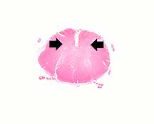
| 02:43, 21 August 2013 | IPLab8Polio1.jpg (file) |  |
3 KB | This is a low-power photomicrograph of a section of spinal cord from this case. Note that the anterior horns (arrows) are almost completely devoid of neurons. | 1 |