File list
This special page shows all uploaded files.
| Date | Name | Thumbnail | Size | Description | Versions |
|---|---|---|---|---|---|
| 21:00, 9 July 2020 | IPLab7Osteosarcoma9b.jpg (file) |  |
361 KB | 1 | |
| 21:00, 9 July 2020 | IPLab7Osteosarcoma8b.jpg (file) |  |
311 KB | 1 | |
| 20:59, 9 July 2020 | IPLab7Osteosarcoma7b.jpg (file) |  |
145 KB | 1 | |
| 21:01, 9 July 2020 | IPLab7Osteosarcoma12b.jpg (file) |  |
353 KB | 1 | |
| 21:00, 9 July 2020 | IPLab7Osteosarcoma11b.jpg (file) |  |
358 KB | 1 | |
| 21:00, 9 July 2020 | IPLab7Osteosarcoma10b.jpg (file) |  |
256 KB | 1 | |
| 01:50, 9 July 2020 | IPLab7Metastatic1a.jpg (file) |  |
30 KB | 1 | |
| 02:12, 9 July 2020 | IPLab7Melanoma7b.jpg (file) |  |
324 KB | 1 | |
| 02:12, 9 July 2020 | IPLab7Melanoma6b.jpg (file) |  |
365 KB | 1 | |
| 02:12, 9 July 2020 | IPLab7Melanoma5b.jpg (file) |  |
365 KB | 1 | |
| 02:11, 9 July 2020 | IPLab7Melanoma4b.jpg (file) |  |
313 KB | 1 | |
| 02:11, 9 July 2020 | IPLab7Melanoma3b.jpg (file) |  |
262 KB | 1 | |
| 02:02, 9 July 2020 | IPLab7IDC1b.jpg (file) |  |
348 KB | 1 | |
| 01:02, 9 July 2020 | IPLab7Fibroadenoma4b.jpg (file) |  |
424 KB | 1 | |
| 01:02, 9 July 2020 | IPLab7Fibroadenoma3b.jpg (file) |  |
459 KB | 1 | |
| 01:02, 9 July 2020 | IPLab7Fibroadenoma2b.jpg (file) |  |
428 KB | 1 | |
| 01:02, 9 July 2020 | IPLab7Fibroadenoma1b.jpg (file) |  |
380 KB | 1 | |
| 01:20, 9 July 2020 | IPLab7ColonCA9a.jpg (file) |  |
101 KB | 1 | |
| 03:25, 9 July 2020 | IPLab7Carcinoid8x.jpg (file) |  |
411 KB | 1 | |
| 03:36, 9 July 2020 | IPLab7Carcinoid4b.jpg (file) |  |
494 KB | 1 | |
| 03:35, 9 July 2020 | IPLab7Carcinoid3b.jpg (file) |  |
563 KB | 1 | |
| 03:25, 9 July 2020 | IPLab7Carcinoid1x.jpg (file) |  |
515 KB | 1 | |
| 03:02, 9 July 2020 | IPLab7Bronchogenic8b.jpg (file) |  |
372 KB | 1 | |
| 02:43, 9 July 2020 | IPLab7Bronchogenic7b.jpg (file) |  |
438 KB | 1 | |
| 02:43, 9 July 2020 | IPLab7Bronchogenic6b.jpg (file) |  |
408 KB | 1 | |
| 03:02, 9 July 2020 | IPLab7Bronchogenic5x.jpg (file) |  |
332 KB | 1 | |
| 02:43, 9 July 2020 | IPLab7Bronchogenic5b.jpg (file) |  |
421 KB | 1 | |
| 00:50, 9 July 2020 | IPLab7Adenoma4b.jpg (file) |  |
324 KB | 1 | |
| 00:50, 9 July 2020 | IPLab7Adenoma3b.jpg (file) |  |
397 KB | 1 | |
| 00:49, 9 July 2020 | IPLab7Adenoma2b.jpg (file) |  |
295 KB | 1 | |
| 00:49, 9 July 2020 | IPLab7Adenoma1b.jpg (file) |  |
358 KB | 1 | |
| 20:14, 20 August 2013 | IPLab6TB6.jpg (file) |  |
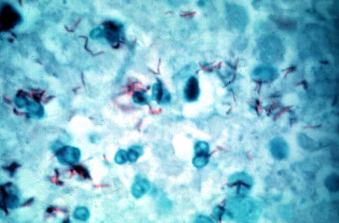
37 KB | This is a high-power (oil immersion) photomicrograph of granuloma stained with an acid-fast stain. Mycobacterium tuberculosis bacilli stain red. | 1 |
| 20:13, 20 August 2013 | IPLab6TB5.jpg (file) |  |
194 KB | High-power photomicrograph of a TB granuloma with multinucleated giant cells adjacent to an area of caseous necrosis (to the left). | 1 |
| 20:11, 20 August 2013 | IPLab6TB4.jpg (file) |  |
68 KB | This is a higher-power photomicrograph of a TB granuloma. The area of caseous necrosis is on the left side of the image, there are multinucleated giant cells and epithelioid macrophages throughout the remainder of the tissue. | 1 |
| 20:10, 20 August 2013 | IPLab6TB3.jpg (file) |  |
72 KB | This is a higher-power photomicrograph of a TB granuloma. Note the eosinophilic material in the center of this granuloma (caseous necrosis) and the epithelioid macrophages and giant cells around the periphery. | 1 |
| 20:10, 20 August 2013 | IPLab6TB2.jpg (file) |  |
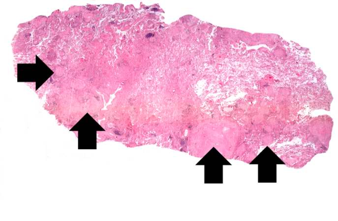
36 KB | This is a low-power photomicrograph of lung tissue with multiple circumscribed nodules - granulomas (arrows). | 1 |
| 20:10, 20 August 2013 | IPLab6TB1.jpg (file) |  |
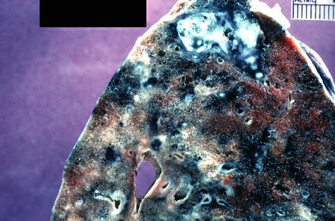
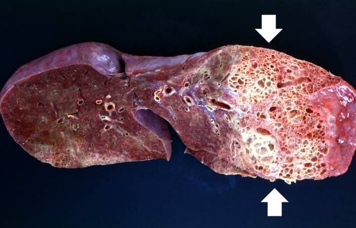
63 KB | This is a photograph of a section of lung with an apical lesion. This lesion represents an old healed lesion from Mycobacterium tuberculosis infection. | 1 |
| 19:59, 20 August 2013 | IPLab6Scleroderma5.jpg (file) |  |
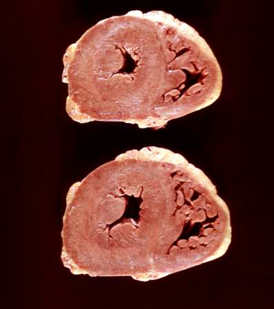
19 KB | This is a gross photograph of the heart from this case. There is thickening of the left ventricular wall and some thickening of the right ventricle as well. | 1 |
| 19:59, 20 August 2013 | IPLab6Scleroderma4.jpg (file) |  |
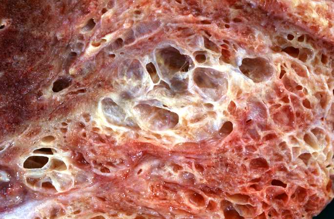
65 KB | This is a closer view of the cut section of lung from this patient showing the extensive fibrosis and the severe emphysematous change. | 1 |
| 19:58, 20 August 2013 | IPLab6Scleroderma3.jpg (file) |  |
64 KB | This is a closer view of the cut section of lung from this patient. Note the extensive fibrosis and the severe emphysematous changes. | 1 |
| 19:58, 20 August 2013 | IPLab6Scleroderma2.jpg (file) |  |
43 KB | This is a gross photograph of a cut section of one lung from this patient. Note the extensive fibrosis lower lobe (arrows). | 1 |
| 19:57, 20 August 2013 | IPLab6Scleroderma1.jpg (file) |  |
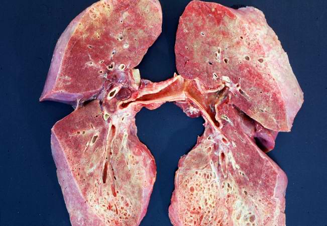
49 KB | This is a gross photograph of cut section of the lungs from this patient. Note the extensive fibrosis of the lung parenchyma. | 1 |
| 20:50, 8 July 2020 | IPLab6RA5b.jpg (file) |  |
407 KB | 1 | |
| 20:50, 8 July 2020 | IPLab6RA3b.jpg (file) |  |
320 KB | 1 | |
| 20:49, 8 July 2020 | IPLab6RA2b.jpg (file) |  |
449 KB | 1 | |
| 17:58, 20 August 2013 | IPLab6PAN9.jpg (file) |  |
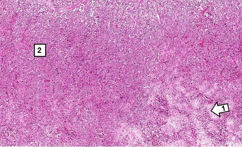
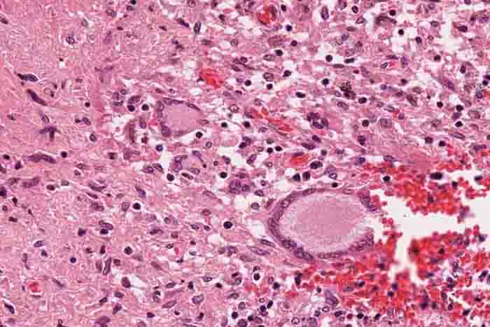
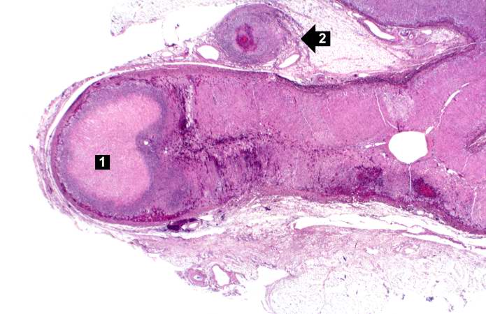
53 KB | This is a low-power photomicrograph of the adrenal gland. There is an area of necrosis in the adrenal (1) and an affected vessel adjacent to the adrenal (2). | 1 |
| 17:58, 20 August 2013 | IPLab6PAN8.jpg (file) |  |
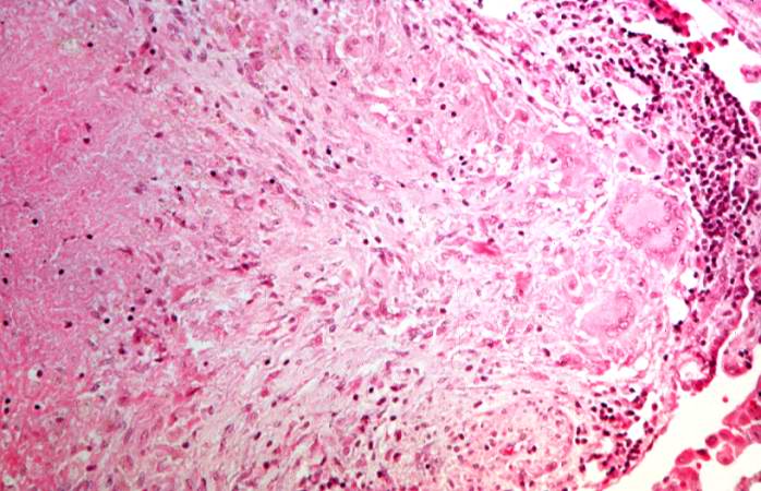
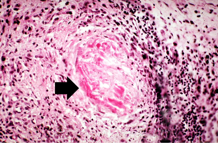
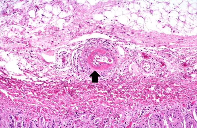
86 KB | This is a high-power photomicrograph of a small vessel with a rim of fibrinoid necrosis (arrow). | 1 |
| 17:57, 20 August 2013 | IPLab6PAN7.jpg (file) |  |
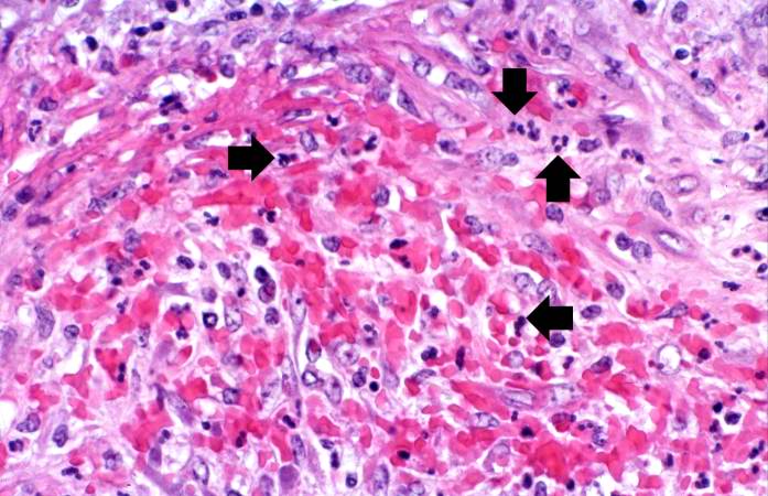
67 KB | This is a high-power photomicrograph of the vessel wall. There is hemorrhage and infiltration with inflammatory cells--primarily neutrophils (arrows). | 1 |
| 17:57, 20 August 2013 | IPLab6PAN6.jpg (file) |  |
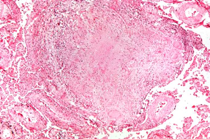
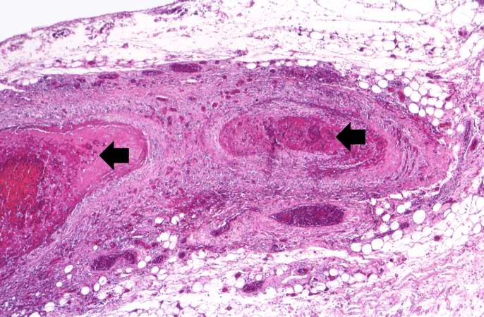
50 KB | his is another example of a mesenteric artery from this case. There is a marked inflammatory cell response surrounding this vessel, fresh hemorrhage (1), and thrombotic material (2). | 1 |
| 17:56, 20 August 2013 | IPLab6PAN5.jpg (file) |  |
79 KB | This is a higher-power photomicrograph of this mesenteric vessel. Note the thrombotic material occluding the vessel (arrows) and the inflammatory cell infiltrate in the wall of the vessel and in the surrounding adventitia. | 1 |