File list
This special page shows all uploaded files.
| Date | Name | Thumbnail | Size | Description | Versions |
|---|---|---|---|---|---|
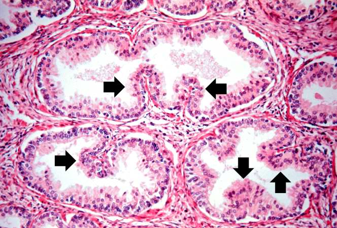
| 15:28, 19 August 2013 | IPLab2Hyperplasia5.jpg (file) |  |
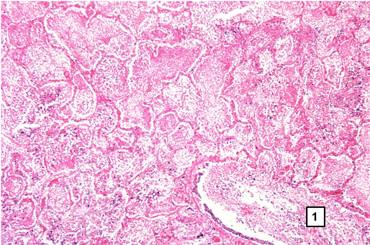
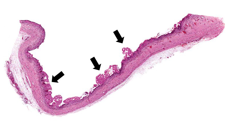
86 KB | Note these glands, which exhibit hyperplasia of the glandular epithelium. The infolding of the glandular epithelial cells forms papillary projections (arrows) into the lumen of the gland. | 1 |
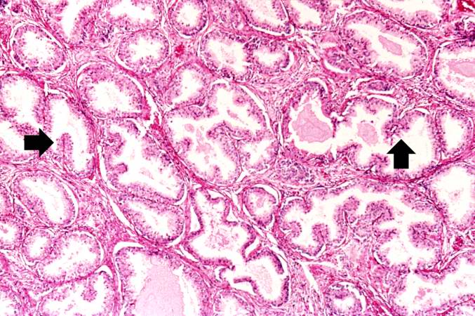
| 15:28, 19 August 2013 | IPLab2Hyperplasia6.jpg (file) |  |
61 KB | Cystic dilatation of glands is present in this photomicrograph. Notice the accumulation of secretory material inside the glands (arrows) and compression (thinning) of the lining epithelium. | 1 |
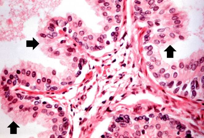
| 15:29, 19 August 2013 | IPLab2Hyperplasia7.jpg (file) |  |
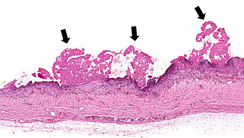
74 KB | A higher-power view shows the papillary folds (arrows) produced by the hyperplastic epithelium projecting into the lumen of the gland. While these papillary folds project into the lumen of the gland, there is no extension through the glandular basement... | 1 |
| 15:29, 19 August 2013 | IPLab2Hyperplasia8.jpg (file) |  |
53 KB | This is a higher-power photomicrograph of papillary folds of hyperplastic epithelium (arrows). | 1 |
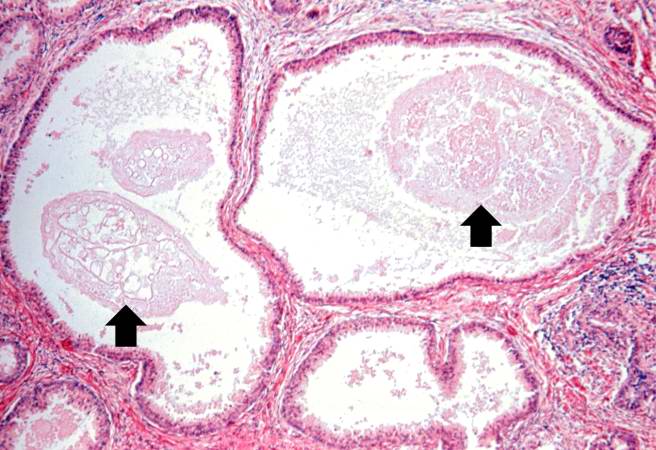
| 15:29, 19 August 2013 | IPLab2Hyperplasia9.jpg (file) |  |
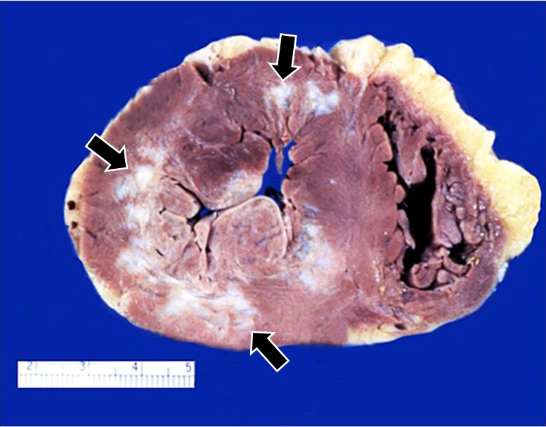
37 KB | This kidney was removed from another autopsy patient who had prostatic hyperplasia resulting in marked urinary retention and back-flow of urine from the bladder into the ureters and renal pelvis. The increased pressure inside the renal pelvis resulted ... | 1 |
| 22:24, 4 September 2013 | IPLab2Hypertrophy2.jpg (file) |  |
204 KB | 2 | |
| 22:26, 4 September 2013 | IPLab2Hypertrophy3.jpg (file) |  |
163 KB | 2 | |
| 22:27, 4 September 2013 | IPLab2Hypertrophy4.jpg (file) |  |
125 KB | 2 | |
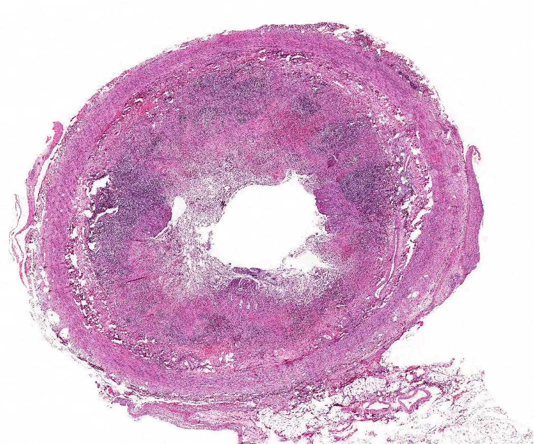
| 15:39, 19 August 2013 | IPLab2Metaplasia1.jpg (file) |  |
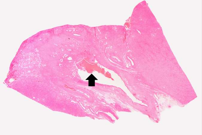
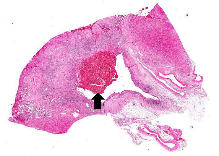
26 KB | This is a low-power photomicrograph showing the full cortical and medullary thickness of the kidney. Note that there is a dilated calyx containing some red blood cells in the center of the section (arrow). The cortex is markedly thin and has severe les... | 1 |
| 21:07, 19 June 2020 | IPLab2Metaplasia1b.JPG (file) |  |
442 KB | 1 | |
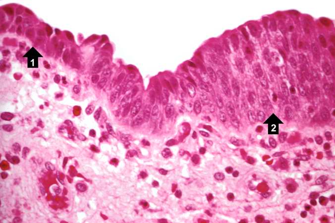
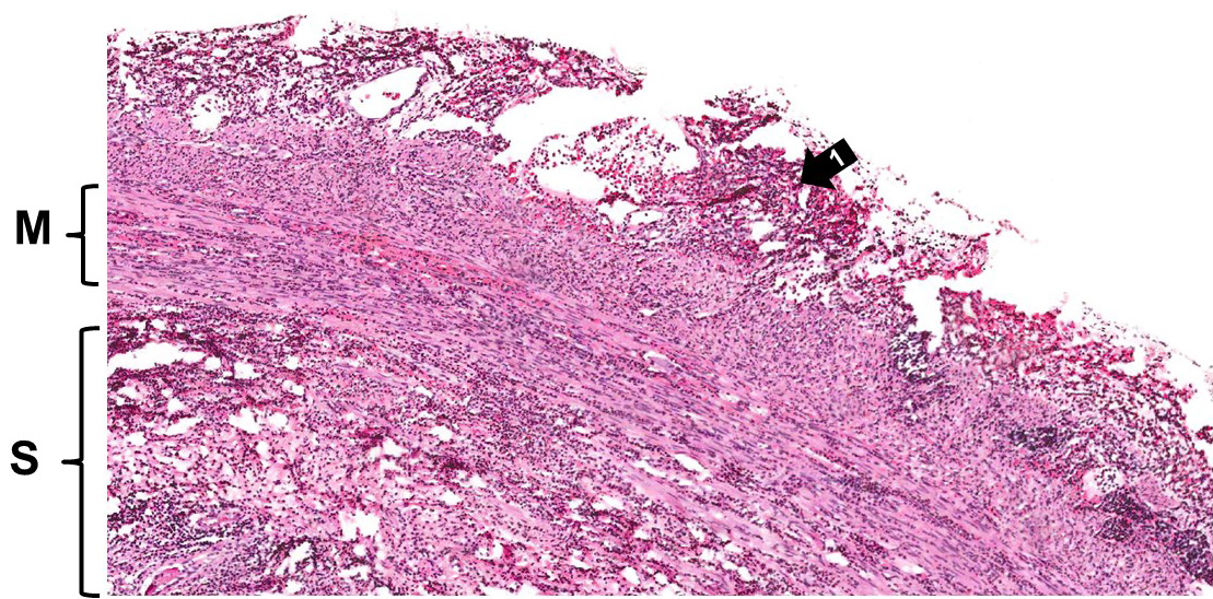
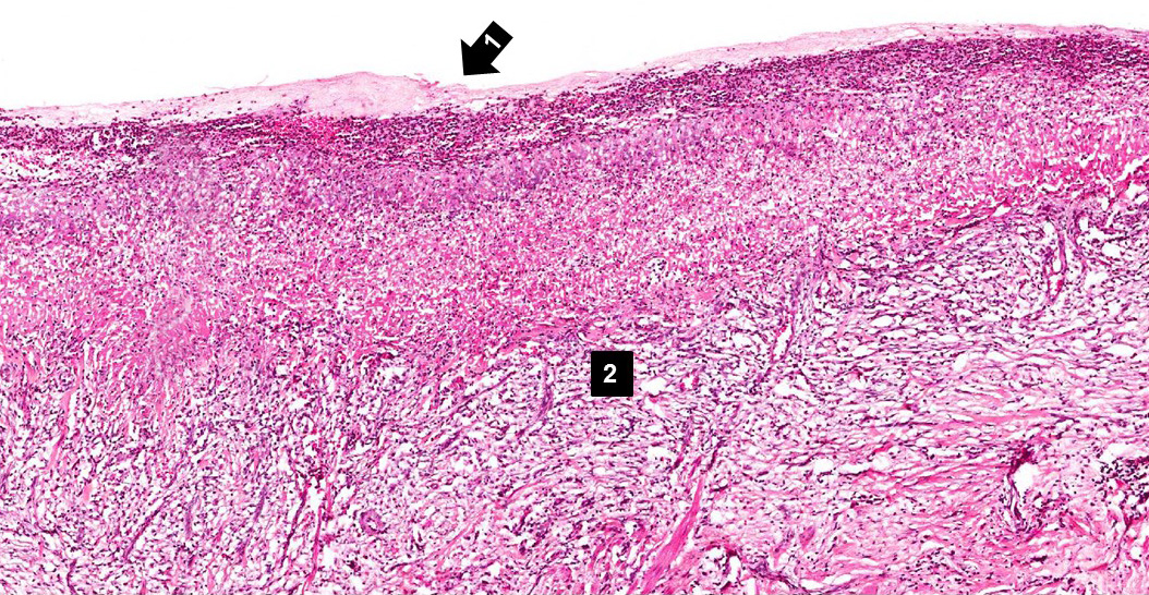
| 15:40, 19 August 2013 | IPLab2Metaplasia2.jpg (file) |  |
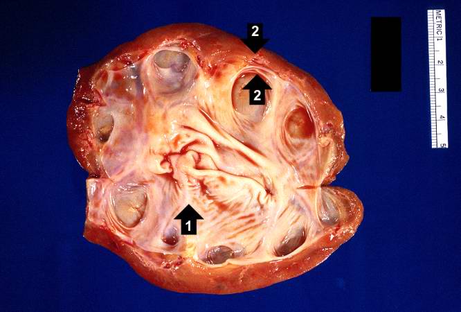
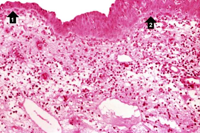
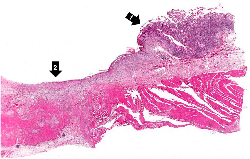
41 KB | his high-power photomicrograph demonstrates the transitional epithelium lining the renal calyx (1) and the junction (transition zone) to a thicker hyperplastic epithelium (2). Note the inflammatory cells and increased vascular response in the stromal t... | 1 |
| 21:07, 19 June 2020 | IPLab2Metaplasia2b.JPG (file) |  |
329 KB | 1 | |
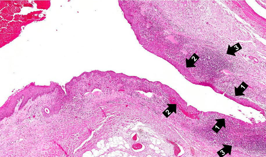
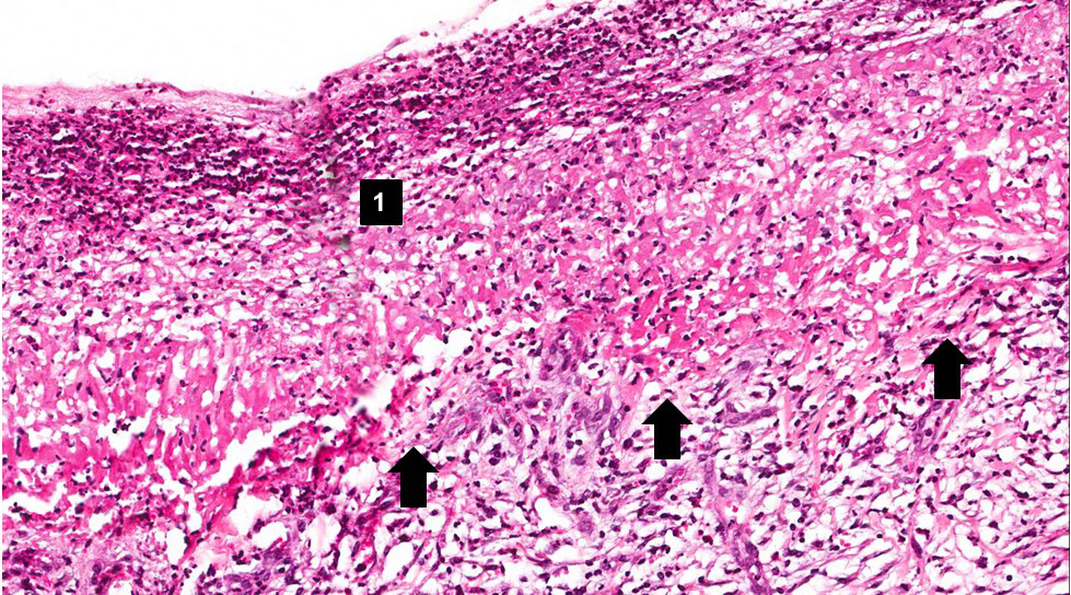
| 15:41, 19 August 2013 | IPLab2Metaplasia3.jpg (file) |  |
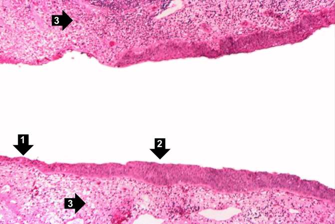
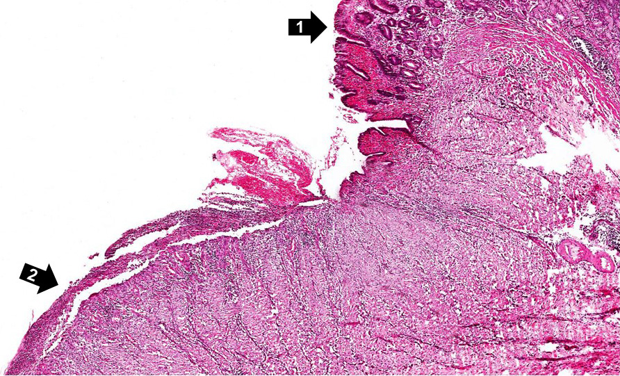
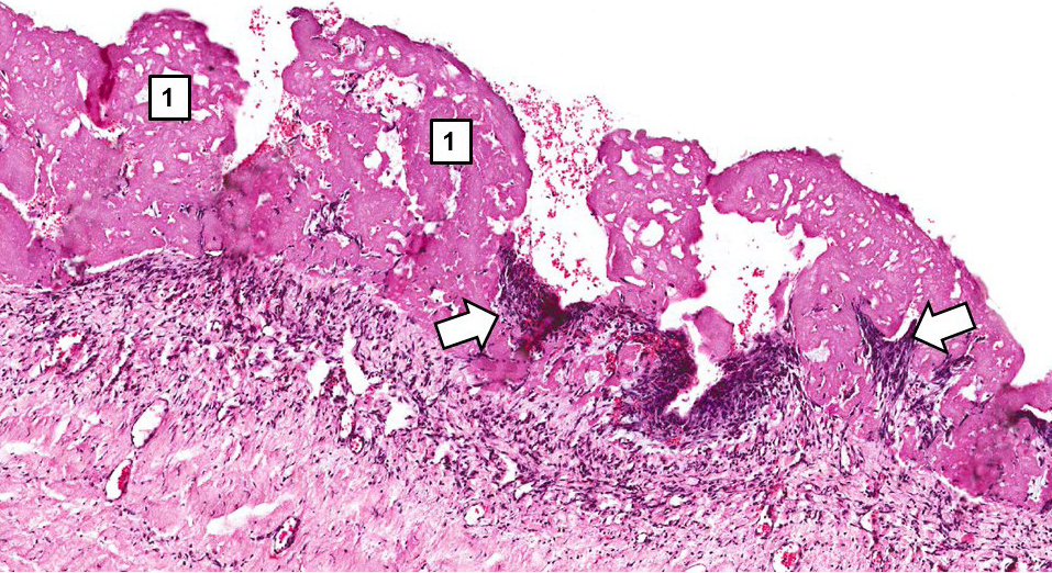
64 KB | A higher-power view shows the junction of normal epithelium (1) with hyperplastic transitional epithelium (2). Note the inflammatory cells in the subepithelial tissue. | 1 |
| 21:08, 19 June 2020 | IPLab2Metaplasia3b.jpg (file) |  |
633 KB | 1 | |
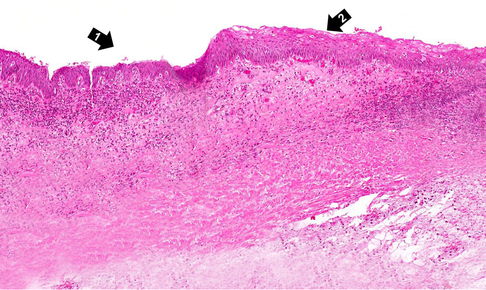
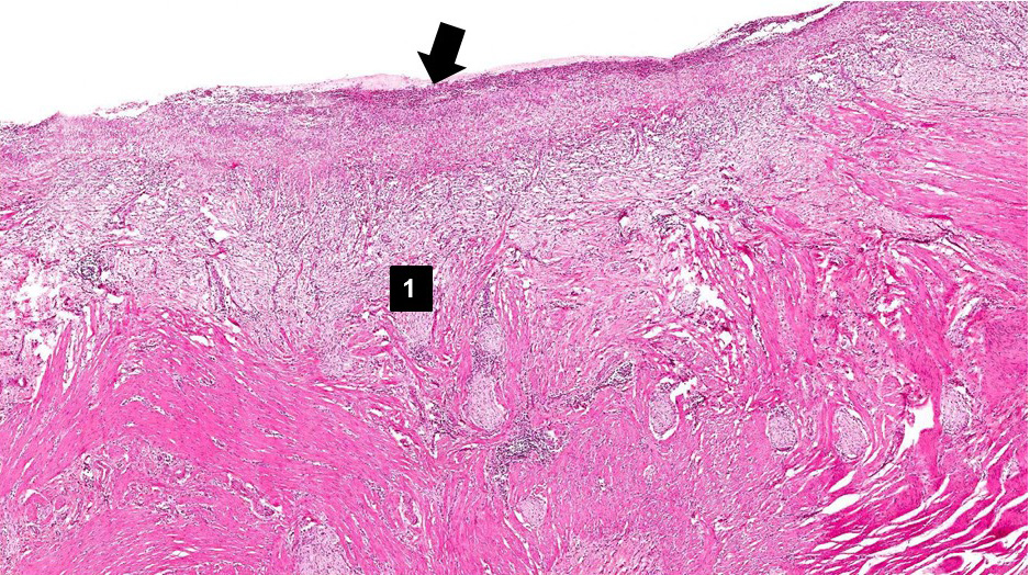
| 15:43, 19 August 2013 | IPLab2Metaplasia4.jpg (file) |  |
46 KB | This is a higher-power photomicrograph of the junction of normal epithelium (1) with hyperplastic transitional epithelium (2). | 1 |
| 21:08, 19 June 2020 | IPLab2Metaplasia4b.jpg (file) |  |
727 KB | 1 | |
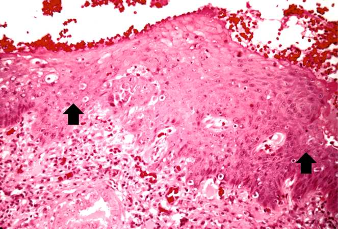
| 15:44, 19 August 2013 | IPLab2Metaplasia5.jpg (file) |  |
76 KB | In areas adjacent to the normal transitional epithelium, there are areas of epithelium (arrows) where the epithelial cells have the character of normal squamous epithelium as found in the dermis. However, squamous epithelium is not normal in the renal ... | 1 |
| 21:08, 19 June 2020 | IPLab2Metaplasia5b.jpg (file) |  |
571 KB | 1 | |
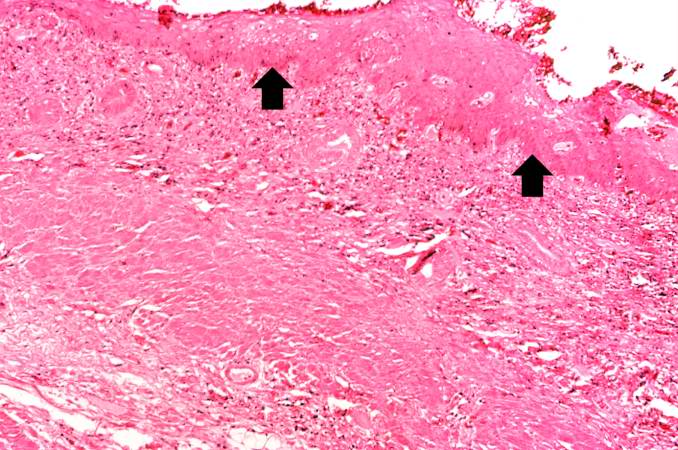
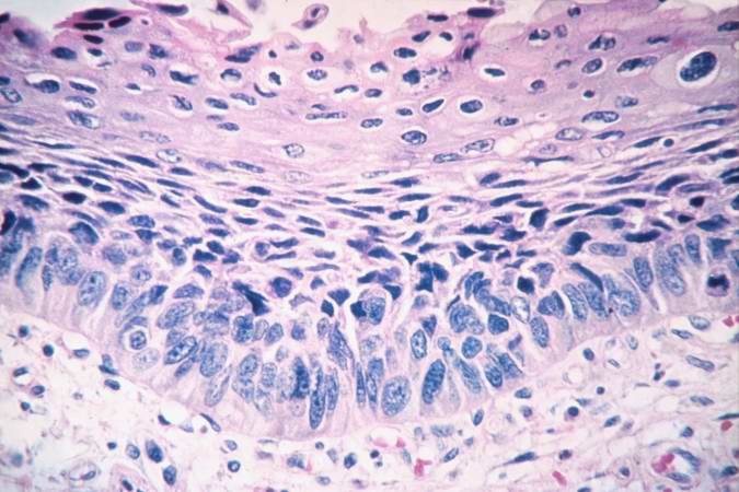
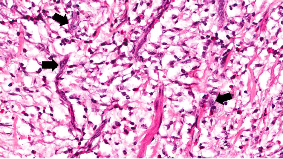
| 15:45, 19 August 2013 | IPLab2Metaplasia6.jpg (file) |  |
67 KB | A high-power photomicrograph of the squamous epithelium shows inflammatory cells in the subepithelial tissue and the formation of keratinized epithelium (arrows). | 1 |
| 15:45, 19 August 2013 | IPLab2Metaplasia7.jpg (file) |  |
57 KB | This is a photomicrograph of the trachea from a smoker. Note that the columnar ciliated epithelium has been replaced by squamous epithelium. | 1 |
| 22:42, 19 June 2020 | IPLab3AcuteAppendicitis2b.jpg (file) |  |
520 KB | 1 | |
| 22:49, 19 June 2020 | IPLab3AcuteAppendicitis2x.jpg (file) |  |
496 KB | 1 | |
| 22:44, 19 June 2020 | IPLab3AcuteAppendicitis3b.jpg (file) |  |
418 KB | 1 | |
| 22:45, 19 June 2020 | IPLab3AcuteAppendicitis4b.jpg (file) |  |
441 KB | 1 | |
| 22:45, 19 June 2020 | IPLab3AcuteAppendicitis5b.jpg (file) |  |
463 KB | 1 | |
| 00:20, 24 June 2020 | IPLab3AcuteMyocardialInfarction1a.JPG (file) |  |
181 KB | 1 | |
| 00:39, 24 June 2020 | IPLab3AcuteMyocardialInfarction1bf.jpg (file) |  |
522 KB | 1 | |
| 01:00, 24 June 2020 | IPLab3AcuteMyocardialInfarction2bs.jpg (file) |  |
651 KB | 1 | |
| 01:02, 24 June 2020 | IPLab3AcuteMyocardialInfarction3b.jpg (file) |  |
800 KB | 1 | |
| 01:02, 24 June 2020 | IPLab3AcuteMyocardialInfarction4b.jpg (file) |  |
584 KB | 1 | |
| 01:03, 24 June 2020 | IPLab3AcuteMyocardialInfarction5b.jpg (file) |  |
587 KB | 1 | |
| 22:22, 23 June 2020 | IPLab3BrainInfarction3b.JPG (file) |  |
249 KB | 1 | |
| 22:23, 23 June 2020 | IPLab3BrainInfarction4b.JPG (file) |  |
395 KB | 1 | |
| 22:23, 23 June 2020 | IPLab3BrainInfarction5b.JPG (file) |  |
346 KB | 1 | |
| 22:23, 23 June 2020 | IPLab3BrainInfarction6b.JPG (file) |  |
351 KB | 1 | |
| 22:24, 23 June 2020 | IPLab3BrainInfarction7b.JPG (file) |  |
239 KB | 1 | |
| 23:06, 19 June 2020 | IPLab3Bronchopneumonia5b.jpg (file) |  |
467 KB | 1 | |
| 23:06, 19 June 2020 | IPLab3Bronchopneumonia6b.jpg (file) |  |
485 KB | 1 | |
| 00:03, 20 June 2020 | IPLab3ChronicPepticUlcer1b.jpg (file) |  |
431 KB | 1 | |
| 00:03, 20 June 2020 | IPLab3ChronicPepticUlcer3b.JPG (file) |  |
459 KB | 1 | |
| 00:04, 20 June 2020 | IPLab3ChronicPepticUlcer4b.JPG (file) |  |
385 KB | 1 | |
| 00:04, 20 June 2020 | IPLab3ChronicPepticUlcer5b.JPG (file) |  |
566 KB | 1 | |
| 00:05, 20 June 2020 | IPLab3ChronicPepticUlcer6b.JPG (file) |  |
483 KB | 1 | |
| 00:04, 20 June 2020 | IPLab3ChronicPepticUlcer7b.JPG (file) |  |
368 KB | 1 | |
| 00:04, 20 June 2020 | IPLab3ChronicPepticUlcer8b.JPG (file) |  |
412 KB | 1 | |
| 23:49, 19 June 2020 | IPLab3FibrinousPericarditis2y.jpg (file) |  |
770 KB | 1 | |
| 23:37, 19 June 2020 | IPLab3FibrinousPericarditis3b.jpg (file) |  |
181 KB | 1 | |
| 23:38, 19 June 2020 | IPLab3FibrinousPericarditis4b.jpg (file) |  |
391 KB | 1 | |
| 23:38, 19 June 2020 | IPLab3FibrinousPericarditis5b.jpg (file) |  |
685 KB | 1 | |
| 01:22, 24 June 2020 | IPLab3HealedMyocardialInfarction1b.jpg (file) |  |
301 KB | 1 |