File list
This special page shows all uploaded files.
| Date | Name | Thumbnail | Size | Description | Versions |
|---|---|---|---|---|---|
| 17:02, 19 August 2013 | IPLab2FattyChange12.jpg (file) |  |
49 KB | This is a cut surface of the same tissue seen in the previous slide. Note the marked nodular pattern. The paler-staining areas between the round nodules represent fibrous connective tissue. | 1 |
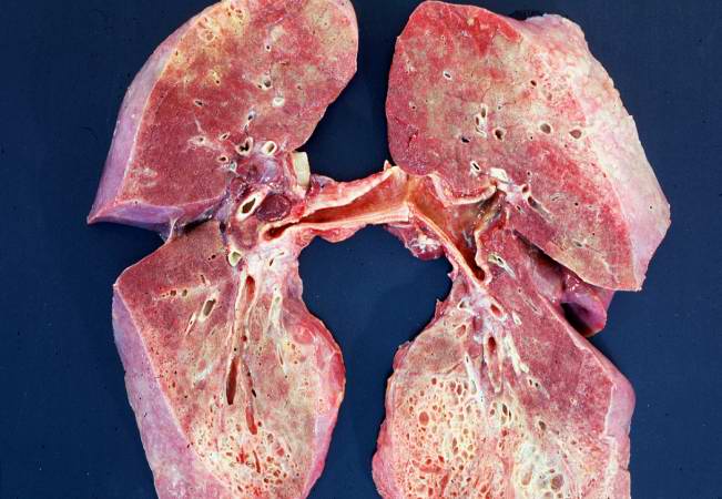
| 19:57, 20 August 2013 | IPLab6Scleroderma1.jpg (file) |  |
49 KB | This is a gross photograph of cut section of the lungs from this patient. Note the extensive fibrosis of the lung parenchyma. | 1 |
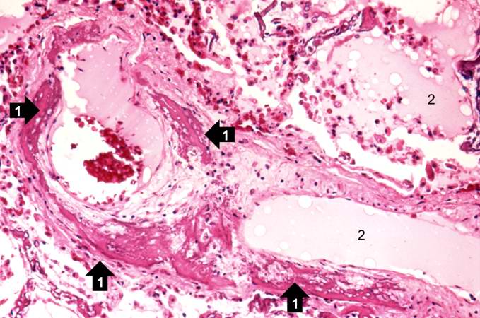
| 17:57, 20 August 2013 | IPLab6PAN6.jpg (file) |  |
50 KB | his is another example of a mesenteric artery from this case. There is a marked inflammatory cell response surrounding this vessel, fresh hemorrhage (1), and thrombotic material (2). | 1 |
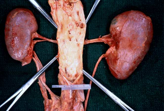
| 16:10, 19 August 2013 | IPLab2Atrophy7.jpg (file) |  |
50 KB | In this gross photograph of kidneys and the abdominal aorta, there is narrowing of the left renal artery at its ostium from the aorta. This atherosclerotic narrowing of the renal artery causes reduced blood pressure in the kidney whose artery is affect... | 1 |
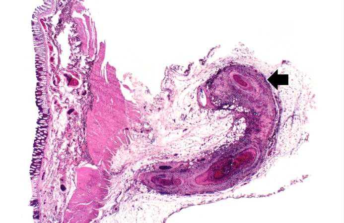
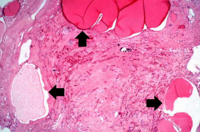
| 17:56, 20 August 2013 | IPLab6PAN4.jpg (file) |  |
51 KB | This is a low-power photomicrograph of a mesenteric vessel from this case of polyarteritis nodosa (arrow). The vessel is completely occluded by thrombotic material and the vessel wall is infiltrated with inflammatory cells. | 1 |
| 16:07, 19 August 2013 | IPLab2Atrophy6.jpg (file) |  |
52 KB | In this slide, the atrophy of the tubules is extending to include the Rete testes (arrow) as well. | 1 |
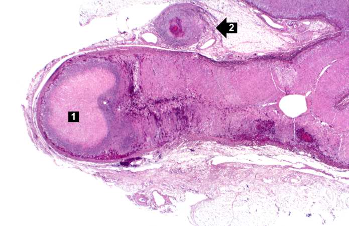
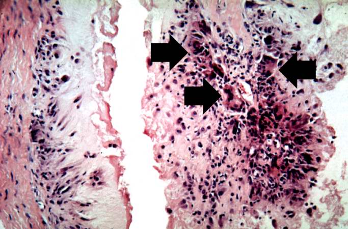
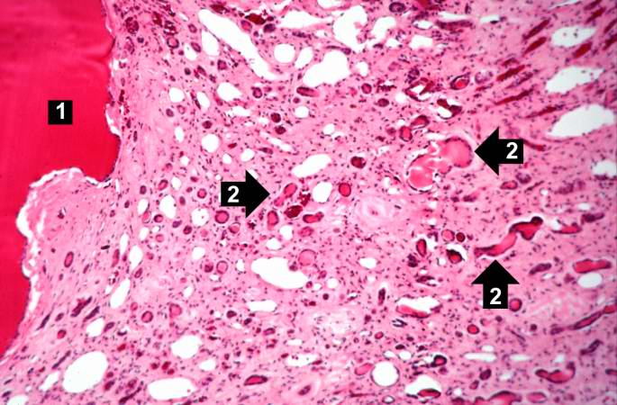
| 17:58, 20 August 2013 | IPLab6PAN9.jpg (file) |  |
53 KB | This is a low-power photomicrograph of the adrenal gland. There is an area of necrosis in the adrenal (1) and an affected vessel adjacent to the adrenal (2). | 1 |
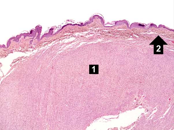
| 17:20, 19 August 2013 | IPLab5Neurofibromatosis5.jpg (file) |  |
53 KB | This is a higher-power photomicrograph of the neurofibroma (1) with the overlying skin (2). | 1 |
| 17:51, 19 August 2013 | IPLab5PolycysticKidney5.jpg (file) |  |
53 KB | This is another low-power photomicrograph of an H&E-stained section from this polycystic kidney. Again note the large cystic structures (arrows)and the fibrous connective tissue throughout this section. | 1 |
| 15:29, 19 August 2013 | IPLab2Hyperplasia8.jpg (file) |  |
53 KB | This is a higher-power photomicrograph of papillary folds of hyperplastic epithelium (arrows). | 1 |
| 15:12, 20 August 2013 | IPLab5Gaucher8.jpg (file) |  |
53 KB | This is a higher-power photomicrograph of the spleen from this case. At this higher power individual cells can be better appreciated and the fibrillar nature of the eosinophilic cytoplasmic material can be seen. | 1 |
| 16:01, 19 August 2013 | IPLab2Atrophy2.jpg (file) |  |
53 KB | This is a low-power photomicrograph of an atrophic testis. Attached to the testis are several vessels (arrow) which are part of the epididymis and the vas deferens. | 1 |
| 17:43, 20 August 2013 | IPLab6Hashimoto3.jpg (file) | 55 KB | This is a higher-power photomicrograph of thyroid from this case. Note the large number of blue-staining inflammatory cells in this tissue. These cells appear to be forming germinal centers. Some residual thyroid gland tissue can be seen in this sectio... | 1 | |
| 14:56, 20 August 2013 | IPLab5Hemochromatosis9.jpg (file) |  |
55 KB | This is a gross picture of pancreas from this case. Note the brown discoloration of the tissue. | 1 |
| 15:28, 20 August 2013 | IPLab5DM6.jpg (file) |  |
56 KB | This is a higher-power photomicrograph of a glomerulus with nodular glomerulosclerosis (arrows). These are the classic Kimmelstiel-Wilson lesions ("K-W lesions") seen in diabetics with nodular glomerulosclerosis. | 1 |
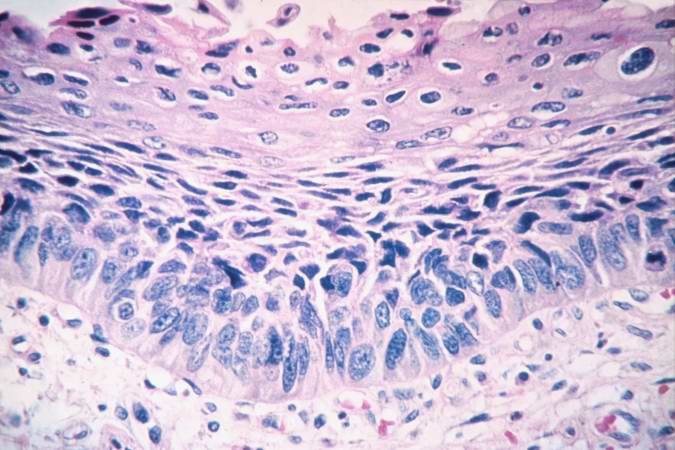
| 15:45, 19 August 2013 | IPLab2Metaplasia7.jpg (file) |  |
57 KB | This is a photomicrograph of the trachea from a smoker. Note that the columnar ciliated epithelium has been replaced by squamous epithelium. | 1 |
| 17:43, 20 August 2013 | IPLab6Hashimoto4.jpg (file) | 58 KB | This is another view of thyroid gland filled with inflammatory cells forming germinal centers (arrows). | 1 | |
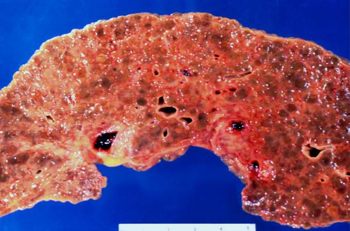
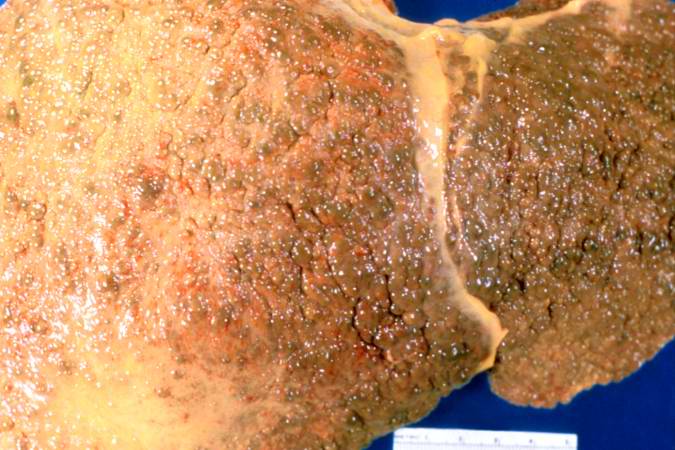
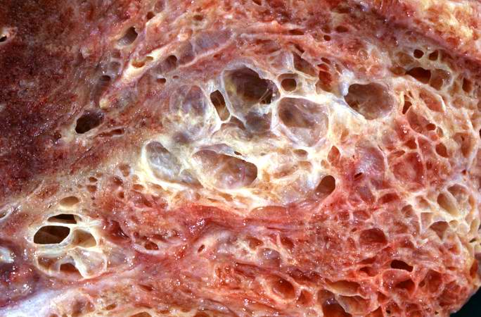
| 17:00, 19 August 2013 | IPLab2FattyChange11.jpg (file) |  |
58 KB | This gross photograph of liver demonstrates severe nodular cirrhosis. Note the extensive scarring of the capsule and the nodular projections of tissue through the uncut capsule in this tissue. The green color is due to the accumulation of bile pigment. | 1 |
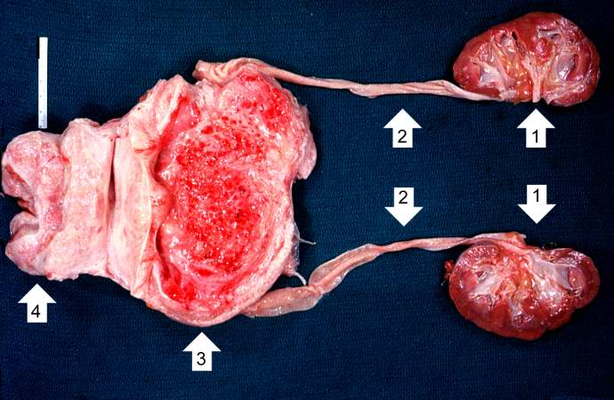
| 15:27, 19 August 2013 | IPLab2Hyperplasia1.jpg (file) |  |
58 KB | This photograph shows the autopsy specimen from this patient. Included are kidneys (1), ureters (2), bladder (3) (which has been opened), and enlarged prostate (4). Note that the bladder mucosa has multiple trabeculae and the bladder mucosa is hyperemi... | 1 |
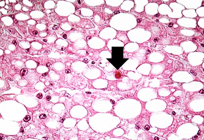
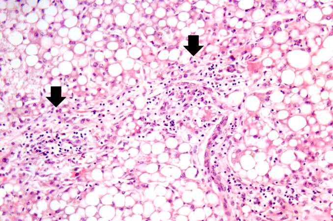
| 16:59, 19 August 2013 | IPLab2FattyChange9.jpg (file) |  |
58 KB | This photomicrograph of the liver is from another patient with a history of alcohol use. There are some clear vacuoles indicating fat droplets (1) and there are numerous red-staining granular deposits within the cytoplasm of hepatocytes (2)--this is al... | 1 |
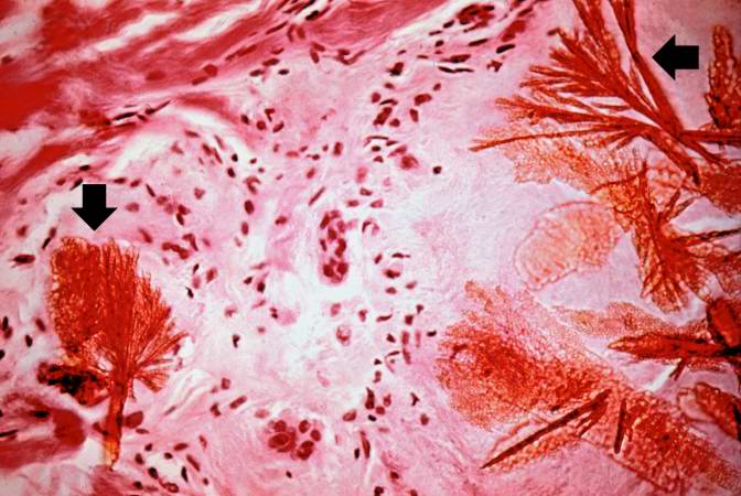
| 15:20, 20 August 2013 | IPLab5Gout7.jpg (file) |  |
59 KB | This is a photomicrograph of a tophus that was fixed in alcohol prior to histologic processing. The alcohol fixation preserves the water soluble urate crystals within the tissue. Note the urate crystals visible in this photomicrograph (arrows). Also no... | 1 |
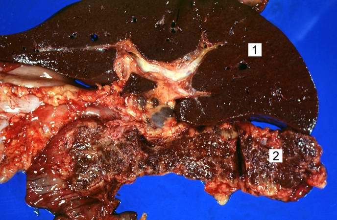
| 14:52, 20 August 2013 | IPLab5Hemochromatosis1.jpg (file) |  |
59 KB | This is a gross photograph of liver (1) and pancreas (2) from this case of hemochromatosis. Note that both of these organs have a dark brown coloration. | 1 |
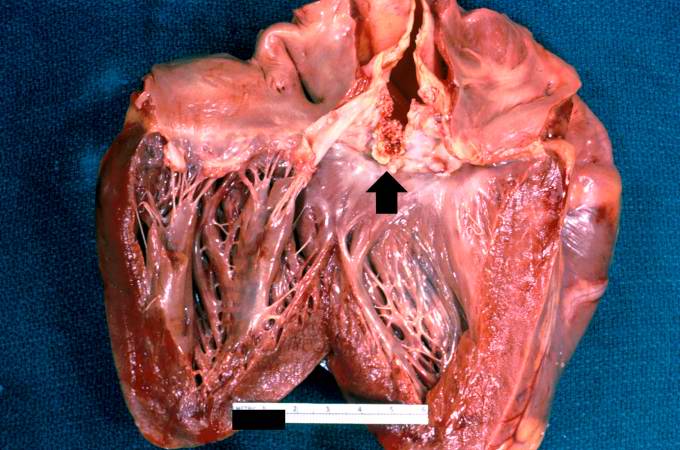
| 16:36, 19 August 2013 | IPLab2Calcification7.jpg (file) |  |
59 KB | A closer view of this same aortic valve (arrow) illustrates the nodularity and thickening of this valve. This valve would be extremely stiff and almost entirely immobile. This particular example of dystrophic calcification is associated with a degenera... | 1 |
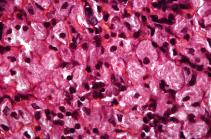
| 17:22, 19 August 2013 | IPLab5Neurofibromatosis8.jpg (file) |  |
59 KB | This is a high-power photomicrograph of the cells in the neurofibroma. | 1 |
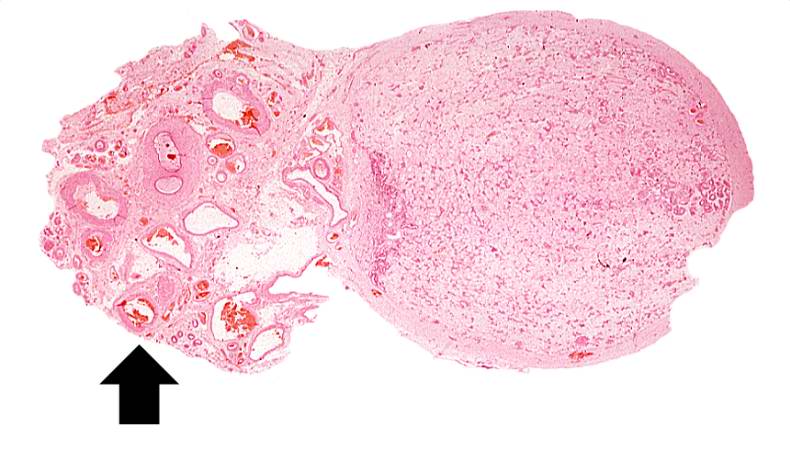
| 15:07, 20 August 2013 | IPLab5Gaucher1.jpg (file) |  |
60 KB | This is a gross photograph of spleen from this case. The spleen is enlarged and the surface is finely granular. | 1 |
| 15:19, 20 August 2013 | IPLab5Gout5.jpg (file) |  |
60 KB | This is a higher-power photomicrograph of the edge of the tophus. Most of the urate crystals dissolve away during processing. The inflammatory cells at the edge of these foci are clearly visible (arrow). | 1 |
| 16:04, 19 August 2013 | IPLab2Atrophy5.jpg (file) |  |
60 KB | A seminiferous tubule is shown on the left containing remnants of spermatocytes (1). There are no mature sperm present. On the right-hand portion of the slide are remnants of other spermatic tubules which have completely atrophied and lost all of their... | 1 |
| 15:19, 20 August 2013 | IPLab5Gout4.jpg (file) |  |
61 KB | This higher-power photomicrograph of the tophus demonstrates the collections of urate crystals (1) and the inflammatory cells at the edge of these foci (2). | 1 |
| 15:28, 19 August 2013 | IPLab2Hyperplasia6.jpg (file) |  |
61 KB | Cystic dilatation of glands is present in this photomicrograph. Notice the accumulation of secretory material inside the glands (arrows) and compression (thinning) of the lining epithelium. | 1 |
| 17:54, 19 August 2013 | IPLab5PolycysticKidney10.jpg (file) |  |
61 KB | This is another photomicrograph of liver demonstrating the histologic appearance of these cysts. These cystic structures are associated with the biliary tree. | 1 |
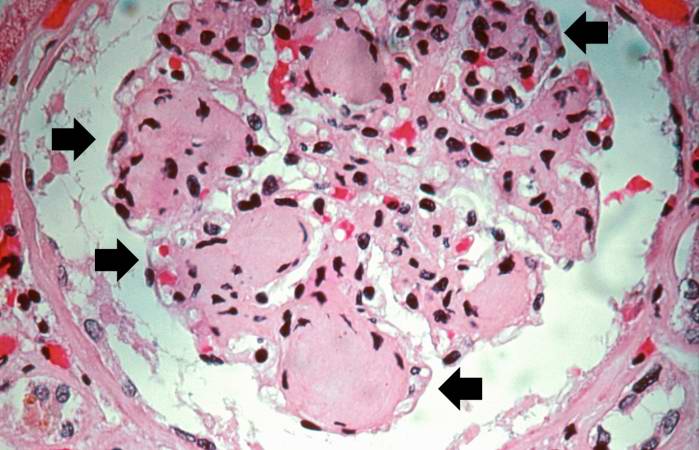
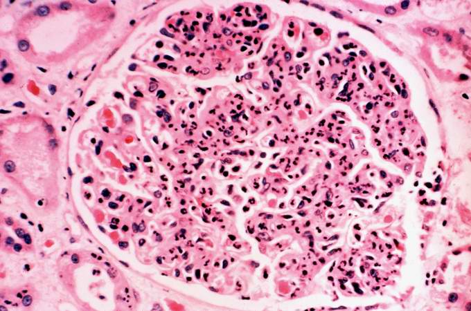
| 20:29, 20 August 2013 | IPLab6GN7.jpg (file) |  |
62 KB | This is a photomicrograph of a glomerulus from another case with acute poststreptococcal glomerulonephritis. In this case the immune complex glomerular disease is ongoing with necrosis and accumulation of neutrophils in the glomerulus. | 1 |
| 15:20, 20 August 2013 | IPLab5Gout6.jpg (file) |  |
63 KB | This is a high-power photomicrograph of the edge of the tophus. The character of the intense chronic inflammatory cell reaction is evident and note the presence of giant cells within this inflammatory cell reaction (arrows). | 1 |
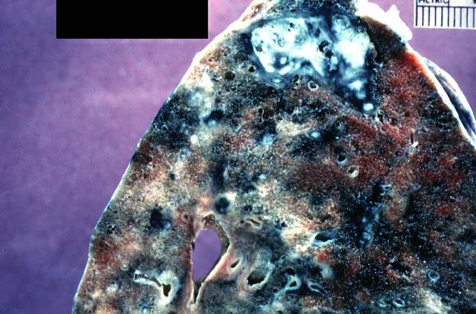
| 20:10, 20 August 2013 | IPLab6TB1.jpg (file) |  |
63 KB | This is a photograph of a section of lung with an apical lesion. This lesion represents an old healed lesion from Mycobacterium tuberculosis infection. | 1 |
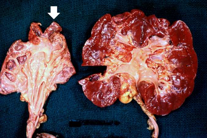
| 16:11, 19 August 2013 | IPLab2Atrophy8.jpg (file) |  |
64 KB | These kidneys were removed from a patient who had blockage of one ureter leading to increased pressure in the renal pelvis. The increased pressure produced hydronephrosis (arrow) in one kidney. What is the cause of atrophy in this case? | 1 |
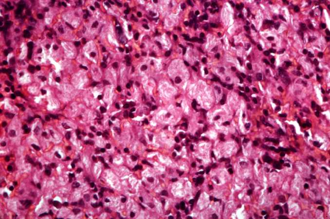
| 15:12, 20 August 2013 | IPLab5Gaucher7.jpg (file) |  |
64 KB | This is another high-power photomicrograph of the spleen from this case. At this high power individual cells can be better appreciated. | 1 |
| 19:58, 20 August 2013 | IPLab6Scleroderma3.jpg (file) |  |
64 KB | This is a closer view of the cut section of lung from this patient. Note the extensive fibrosis and the severe emphysematous changes. | 1 |
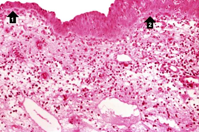
| 15:41, 19 August 2013 | IPLab2Metaplasia3.jpg (file) |  |
64 KB | A higher-power view shows the junction of normal epithelium (1) with hyperplastic transitional epithelium (2). Note the inflammatory cells in the subepithelial tissue. | 1 |
| 16:01, 19 August 2013 | IPLab2Atrophy3.jpg (file) |  |
65 KB | This is a higher-power photomicrograph of an atrophic testis. In this section there are seminiferous tubules with viable cells (1) although there are no visible spermatocytes. Other seminiferous tubules are completely acellular and have a pale pink hya... | 1 |
| 19:59, 20 August 2013 | IPLab6Scleroderma4.jpg (file) |  |
65 KB | This is a closer view of the cut section of lung from this patient showing the extensive fibrosis and the severe emphysematous change. | 1 |
| 16:58, 19 August 2013 | IPLab2FattyChange7.jpg (file) |  |
65 KB | A high-power photomicrograph of the liver parenchyma shows that each individual liver cell is filled with a large, clear droplet which represents the space remaining after lipid was dissolved by the dehydration procedure used to embed the tissue. '''No... | 1 |
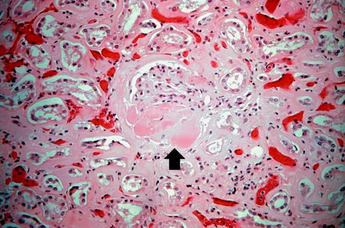
| 16:34, 19 August 2013 | IPLab2Calcification4.jpg (file) |  |
65 KB | This high-power photomicrograph of a blood vessel shows calcium deposits in the vascular wall (1) and proteinaceous material (2) (from edema) within some of the alveoli. The smooth muscle in the vessel wall has been almost completely replaced by calciu... | 1 |
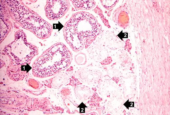
| 17:52, 19 August 2013 | IPLab5PolycysticKidney6.jpg (file) |  |
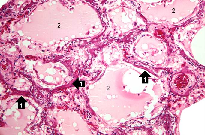
66 KB | This is a higher-power photomicrograph of polycystic kidney showing the edge of a large cyst (1). In this section there are numerous tubules and dilated collecting ducts (2) that are filled with the same red proteinaceous material as the larger cysts. | 1 |
| 17:52, 19 August 2013 | IPLab5PolycysticKidney7.jpg (file) |  |
66 KB | This high-power photomicrograph shows abnormal glomeruli (arrows) and some tubules. | 1 |
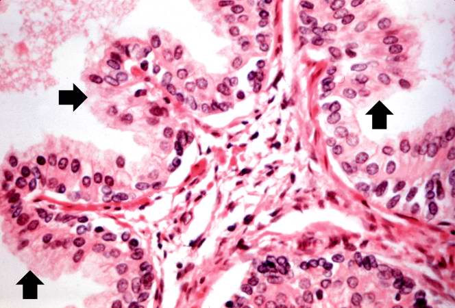
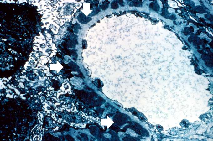
| 20:29, 20 August 2013 | IPLab6GN6.jpg (file) |  |
66 KB | This is an electron micrograph of subepithelial granular electron dense deposits (arrows) which correspond to the granular immunofluorescence seen in the previous image. | 1 |
| 15:29, 20 August 2013 | IPLab5DM7.jpg (file) |  |
66 KB | This is a photomicrograph of kidney with a focal exudative lesion in a glomerulus (arrow) and sclerosis, interstitial fibrosis, and congestion. | 1 |
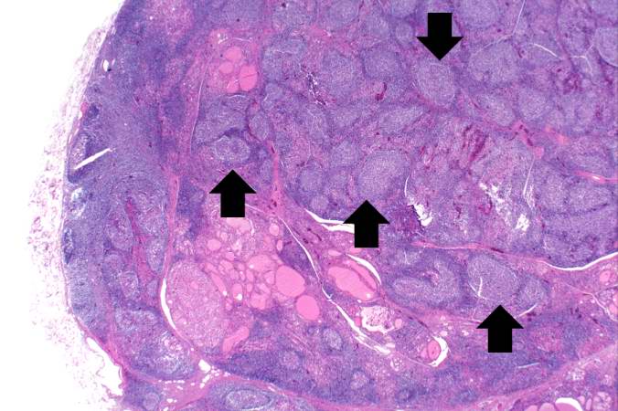
| 16:57, 19 August 2013 | IPLab2FattyChange6.jpg (file) |  |
67 KB | Another view at the same power illustrates the proliferation of bile ducts in the interlobular and perichordal regions (arrows). | 1 |
| 15:45, 19 August 2013 | IPLab2Metaplasia6.jpg (file) |  |
67 KB | A high-power photomicrograph of the squamous epithelium shows inflammatory cells in the subepithelial tissue and the formation of keratinized epithelium (arrows). | 1 |
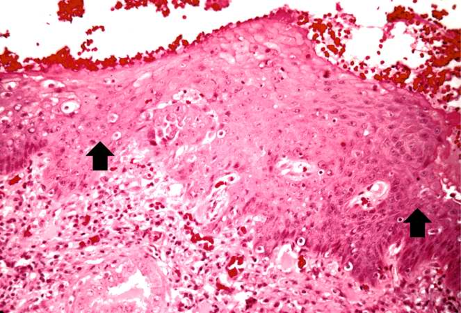
| 18:13, 19 August 2013 | IPLab5Antitrypsin3.jpg (file) |  |
67 KB | This is a gross photograph of the bronchi and lungs. Note the hemorrhage in the bronchi and in the lung parenchyma. | 1 |
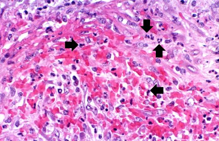
| 17:57, 20 August 2013 | IPLab6PAN7.jpg (file) |  |
67 KB | This is a high-power photomicrograph of the vessel wall. There is hemorrhage and infiltration with inflammatory cells--primarily neutrophils (arrows). | 1 |
| 16:34, 19 August 2013 | IPLab2Calcification5.jpg (file) |  |
68 KB | This photomicrograph demonstrates pulmonary alveoli with extensive calcium depositions (1) in the septa and protein accumulations (2) in the alveoli. | 1 |