File list
This special page shows all uploaded files.
| Date | Name | Thumbnail | Size | Description | Versions |
|---|---|---|---|---|---|
| 05:28, 21 August 2013 | IPLab12RadiationChanges2.jpg (file) |  |
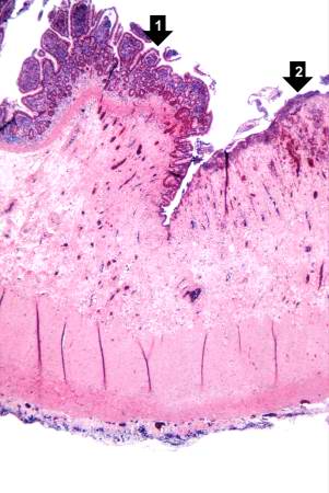
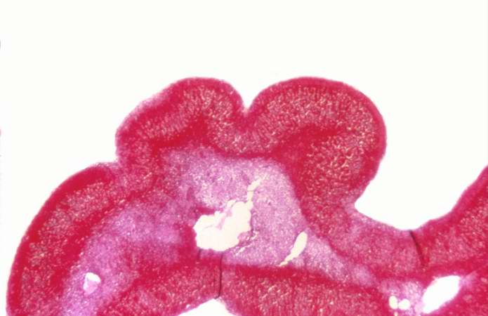
26 KB | This is a higher-power photomicrograph of the surgical specimen of the ileum showing the transition from the normal epithelium (1) to the atrophied epithelium (2) in the area of radiation injury. | 1 |
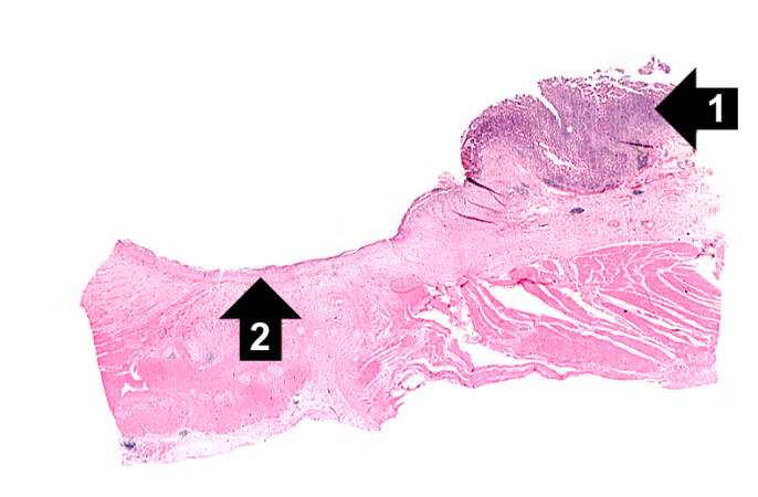
| 05:27, 21 August 2013 | IPLab12RadiationChanges1.jpg (file) |  |
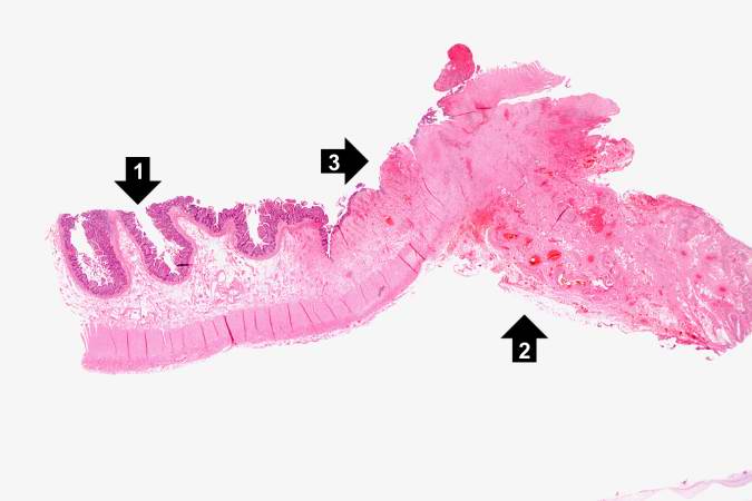
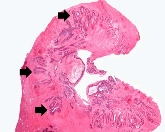
26 KB | This is a low-power photomicrograph of the surgical specimen of the ileum. The normal ileum is to the left (1). The area of stricture consists of dense fibrous connective tissue (2) and there is loss or marked atrophy of the epithelium (3). | 1 |
| 05:23, 21 August 2013 | IPLab12RadiationFibrosis2.jpg (file) |  |
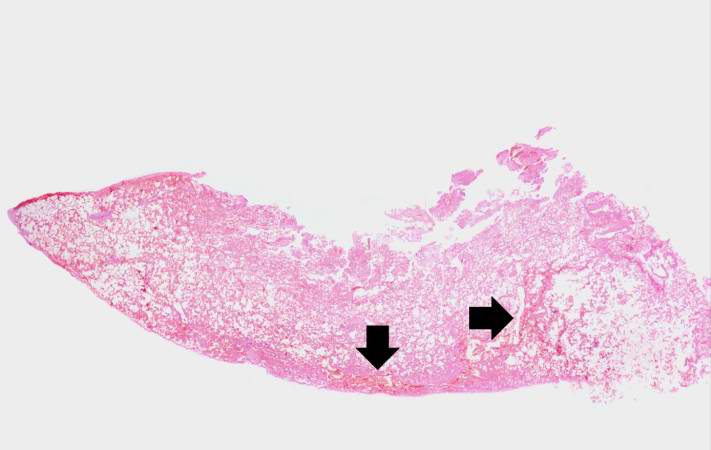
27 KB | This is a gross photograph of cut sections of lung. There are several areas of fibrosis (arrows) within the lung parenchyma. | 1 |
| 02:37, 21 August 2013 | IPLab8HSVEncephalitis11.jpg (file) |  |
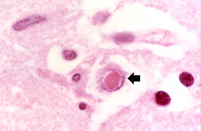
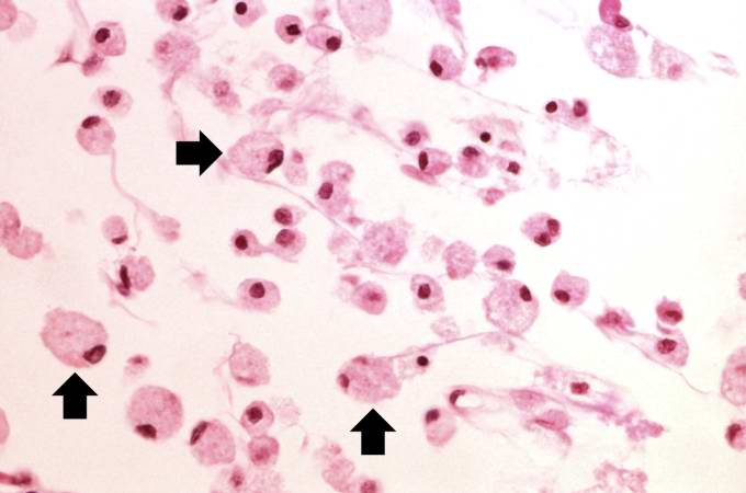
27 KB | This is another high-power photomicrograph of a cell containing an intranuclear inclusion body (arrow). | 1 |
| 05:44, 21 August 2013 | IPLab13Myelomeningocele1.jpg (file) |  |
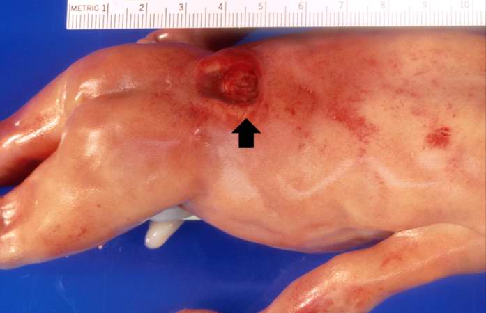
27 KB | This is a gross photograph of the fetus at autopsy. Note the defect in the lower lumbar region of the spinal column (arrow). The myelomeningocele can be seen protruding from this defect. | 1 |
| 03:42, 16 August 2013 | IPLab1Prostate6.jpg (file) |  |
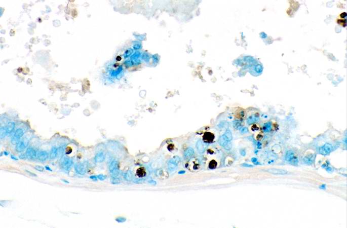
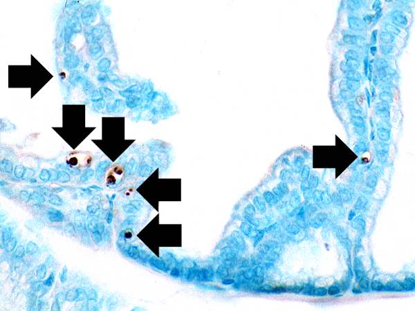
27 KB | This is a higher-power photomicrograph of prostatic epithelium with the TUNEL staining. Note the apoptotic cells (brown nuclei) in the epithelium as well as those floating freely. | 1 |
| 03:49, 21 August 2013 | IPLab9Diphtheria1.jpg (file) |  |
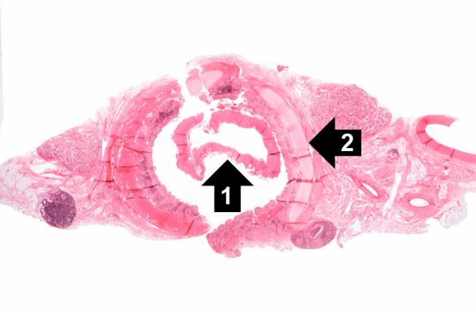
28 KB | This is a low-power photomicrograph of the trachea with the diphtheritic membrane (1), which has pulled away from the tracheal lining during histological processing. Note the tracheal cartilage (2) present in this section. | 1 |
| 02:14, 21 August 2013 | IPLab7Osteosarcoma6.jpg (file) |  |
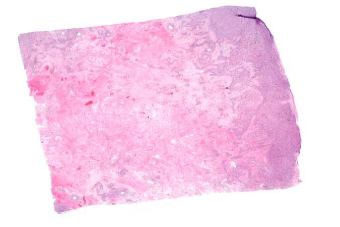
28 KB | This is a low-power photomicrograph of decalcified histologic section from this tumor. Note the blue color (cell nuclei stain blue) of much of this section indicating the increased cellularity of the tumor. | 1 |
| 03:22, 19 August 2013 | IPLab3Bronchopneumonia3.jpg (file) |  |
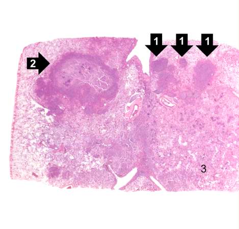
28 KB | This is a low-power photomicrograph of lung with multiple focal lesions (1) throughout the tissue, some of which have a pale center indicating a loss of parenchymal tissue (2). This is typical of abscess formation in the lung and represents a form of l... | 1 |
| 04:23, 19 August 2013 | IPLab3BrainInfarction8.jpg (file) |  |
28 KB | This is a high-power photomicrograph of gitter cells (arrows). | 1 |
| 04:10, 19 August 2013 | IPLab3FibrinousPericarditis4.jpg (file) |  |
28 KB | This is a higher-power photomicrograph demonstrating fronds of fibrin (arrows) projecting from the surface of the pericardium. | 1 |
| 03:52, 21 August 2013 | IPLab9ARF2.jpg (file) |  |
29 KB | This is a low-power photomicrograph of heart tissue. Little can be seen at this magnification, except that the tissue looks relatively normal. | 1 |
| 05:01, 21 August 2013 | IPLab11Ascariasis6.jpg (file) |  |
29 KB | This high-power photomicrograph of the fecal specimen from this patient shows a Giardia lamblia trophozoite. Note the two nuclei and the tapered end (that goes out of the plane of focus) containing flagella (arrow). | 1 |
| 03:47, 19 August 2013 | IPLab3ForeignBodyGranuloma1.jpg (file) |  |
29 KB | This is a low-power photomicrograph of lung and pleura. There is some hemorrhage in this tissue (arrows), probably the result of surgery or the gunshot wounds. | 1 |
| 18:22, 19 August 2013 | IPLab6GravesDisease2.jpg (file) |  |
29 KB | This is a low-power photomicrograph of thyroid tissue from this case. The tissue is very cellular with very little colloid. | 1 |
| 06:04, 21 August 2013 | IPLab13Meningococcemia1.jpg (file) |  |
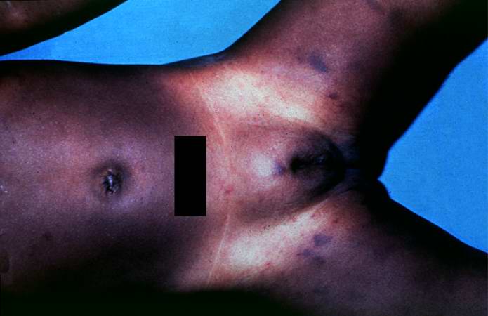
29 KB | In this gross photograph from the autopsy, note the areas of hemorrhage in the inguinal region. | 1 |
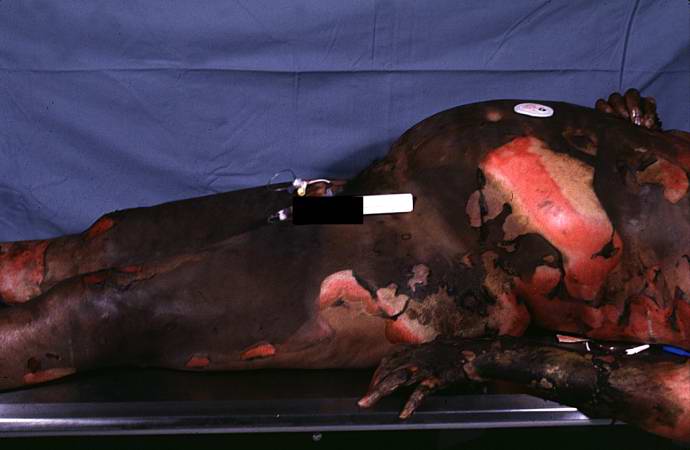
| 05:37, 21 August 2013 | IPLab12Burns1.jpg (file) |  |
30 KB | This photograph taken at autopsy demonstrates the severity of the surface burns on this patient. | 1 |
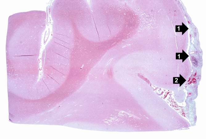
| 03:46, 21 August 2013 | IPLab9BacterialMeningitis2.jpg (file) |  |
30 KB | This is a low-power photomicrograph of brain section. Note the exudate (1) in the meninges and congestion of the vessels (2) in the leptomeninges. | 1 |
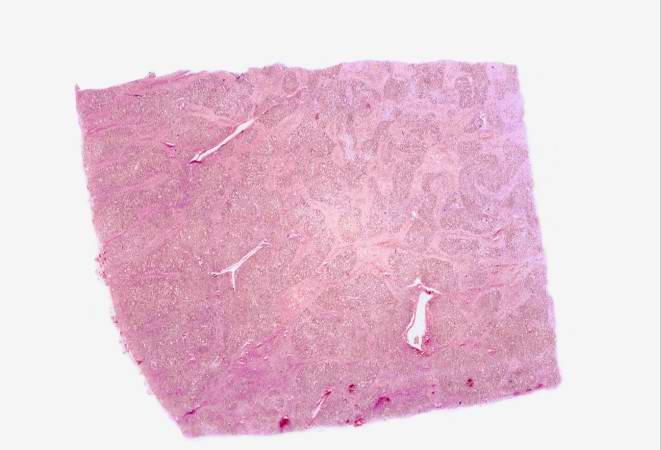
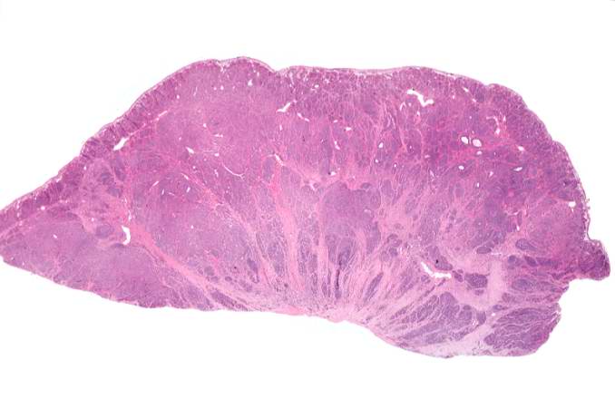
| 05:47, 21 August 2013 | IPLab13BiliaryAtresia1.jpg (file) |  |
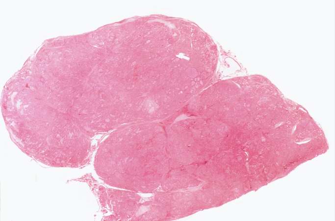
30 KB | This is a low power photomicrograph of a section of liver. Even at this low magnification, areas of fibrosis can be appreciated. | 1 |
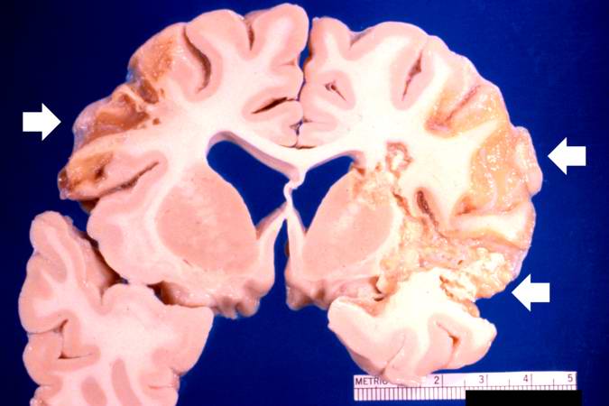
| 05:17, 21 August 2013 | IPLab12Alcoholic14.jpg (file) |  |
30 KB | This is another photograph of the cerebellum from this patient demonstrating the marked thinning of the anterior portion of the cerebellum (arrows). This pattern of cerebellar damage is consistent with Wernicke's encephalopathy. | 1 |
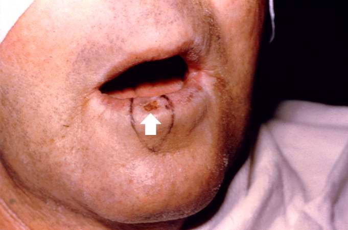
| 01:31, 21 August 2013 | IPLab7LipSCC1.jpg (file) |  |
30 KB | This is a pre-op photograph of this patient with an ulcerated lesion on his lip (arrow). Also note that the lip is somewhat thickened. The area for surgical excision is delineated by black marker. | 1 |
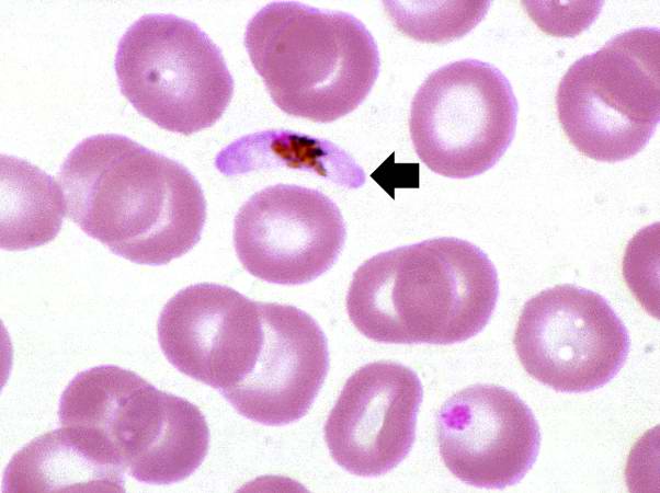
| 04:55, 21 August 2013 | IPLab11Malaria4.jpg (file) |  |
30 KB | In this high power photomicrograph of a thin smear of blood from this patient there is one P. falciparum gametocyte (arrow). These gametocytes have a characteristic "banana" shape. | 1 |
| 16:14, 15 August 2013 | IPLab1LungAbscess1.jpg (file) |  |
30 KB | 1 | |
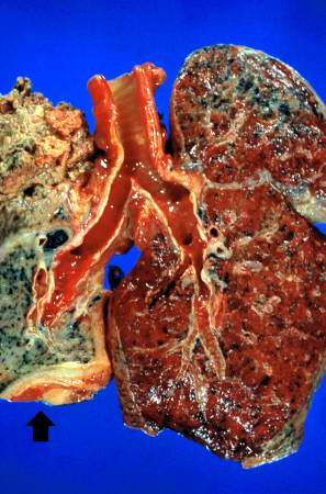
| 05:32, 21 August 2013 | IPLab12Mesothelioma2.jpg (file) |  |
30 KB | This is a gross photograph of cut sections of the lungs. The right lung is congested. The left lung is shrunken and filled with tumor. There is a thick layer of tumor along the inferior portion of the lung (arrow). | 1 |
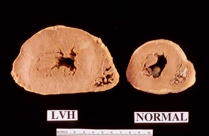
| 23:29, 18 August 2013 | IPLab2Hypertrophy1.jpg (file) |  |
30 KB | This is a gross photograph of a cross section of a normal human heart taken at autopsy (right) and the heart from this case, which demonstrates concentric hypertrophy of the left ventricular wall. Note the marked thickening of the left ventricular wall... | 1 |
| 05:55, 21 August 2013 | IPLab13WT4.jpg (file) |  |
30 KB | This lowest-power view shows the tumor itself; no tissue is present that can be readily identified as normal kidney. There does appear to be a capsule surrounding the tumor. Eosinophilic bands are seen surrounding basophilic islands of cells. These cor... | 1 |
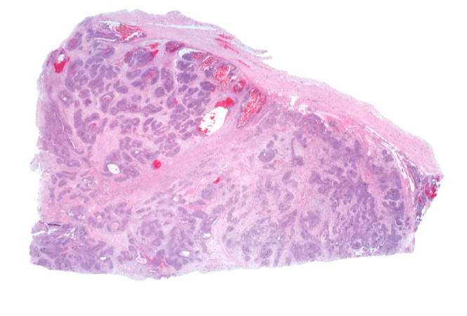
| 02:06, 21 August 2013 | IPLab7Carcinoid1.jpg (file) |  |
31 KB | This is a low-power photomicrograph of the surgical specimen showing basophilic and eosinophilic areas delimiting areas of tumor infiltration. | 1 |
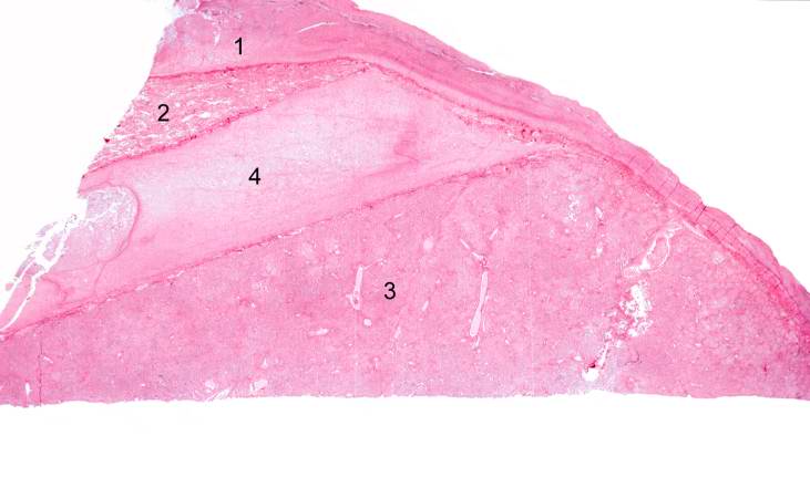
| 03:17, 19 August 2013 | IPLab3LobarPneumonia4.jpg (file) |  |
31 KB | This low-power photomicrograph of the lung shows: (1) markedly thickened pleura, indicating an inflammatory process which has been present for several days; (2) a small wedge-shaped segment of normal lung which is somewhat compressed due to artifact; (... | 1 |
| 04:15, 19 August 2013 | IPLab3ChronicPepticUlcer3.jpg (file) |  |
31 KB | This is a low-power photomicrograph of the transected ulcer. The blue cells on the right hand side of this section are the normal gastric epithelial cells of the mucosa (1). Note the absence of any epithelial cells within the crater of the ulcer (2). | 1 |
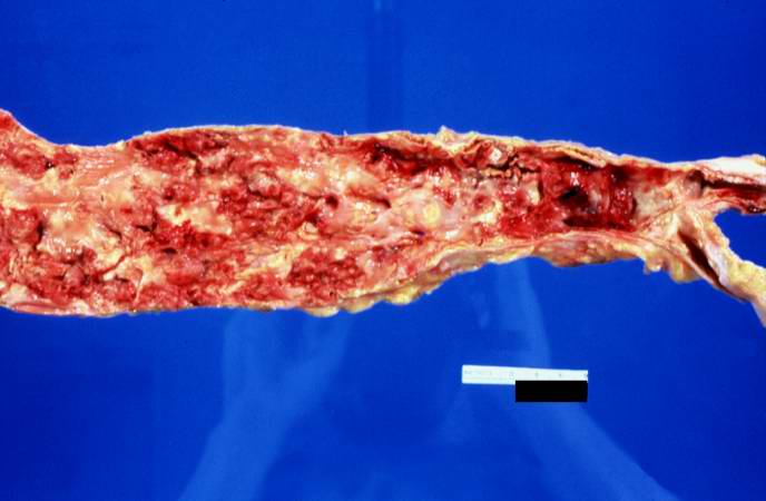
| 17:00, 19 August 2013 | IPLab4AtheromatousEmboli1.jpg (file) |  |
31 KB | This is a gross photograph of the aorta from this patient opened lengthwise with the luminal surface visible. Note the rough surface with ulcerations and adherent thrombotic material. There is a mild dilation (aneurysm) at the distal aorta just at the ... | 1 |
| 05:20, 21 August 2013 | IPLab12Acetaminophen6.jpg (file) |  |
32 KB | Photograph taken at autopsy demonstrating the severe necrosis of the skin of foot. | 1 |
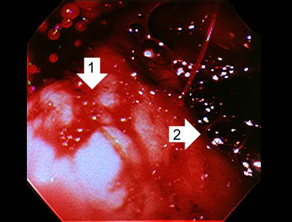
| 05:15, 21 August 2013 | IPLab12Alcoholic7.jpg (file) |  |
32 KB | This photograph was taken during the EGD while the patient was alive. Note the red hyperemic areas (1) and the area of hemorrhage (2). | 1 |
| 02:34, 21 August 2013 | IPLab8HSVEncephalitis1.jpg (file) |  |
32 KB | This is a gross photograph of a section of brain showing multiple small, punctate hemorrhages throughout the brain parenchyma (arrows). | 1 |
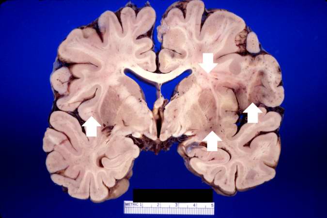
| 04:22, 19 August 2013 | IPLab3BrainInfarction2.jpg (file) |  |
32 KB | This is a gross photograph of a cross-section of brain demonstrating the areas of infarction (arrows). | 1 |
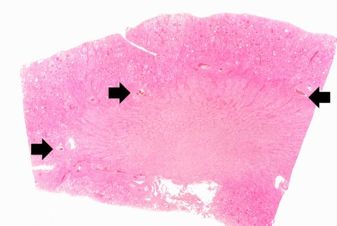
| 17:00, 19 August 2013 | IPLab4AtheromatousEmboli3.jpg (file) |  |
34 KB | This is a low-power photomicrograph of kidney tissue. Several blood vessels can be identified at the corticomedullary junction (arrows). | 1 |
| 06:05, 21 August 2013 | IPLab13Meningococcemia7.jpg (file) |  |
34 KB | This higher-power photomicrograph of the adrenal gland from this case provides an example of hemorrhagic necrosis. | 1 |
| 04:07, 21 August 2013 | IPLab10Histo7.jpg (file) |  |
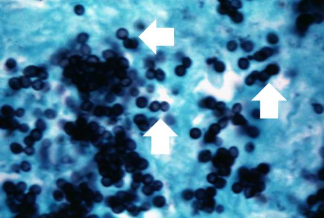
34 KB | This photomicrograph was taken under oil immersion to show the silver-stained Histoplasma organisms. Some of the organisms appear to be budding (arrows). | 1 |
| 01:36, 21 August 2013 | IPLab7EsophSCC2.jpg (file) |  |
34 KB | This low-power photomicrograph of a cross-section through the esophagus at the area of constriction shows extensive infiltration of the esophageal wall with squamous cell carcinoma (arrows). | 1 |
| 05:39, 21 August 2013 | IPLab12Burns9.jpg (file) |  |
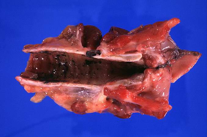
34 KB | This photograph demonstrates the black carbonaceous material in the trachea. | 1 |
| 05:37, 21 August 2013 | IPLab12Burns2.jpg (file) |  |
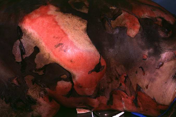
34 KB | This closer view shows the burned skin peeling off to expose the underlying tissue. The various depths of the burn can be appreciated by the color and character of the underlying tissue. | 1 |
| 05:07, 21 August 2013 | IPLab11Chagas2.jpg (file) |  |
34 KB | This peripheral blood smear from the patient shows a higher power view of a Trypanosoma cruzi trypomastigote. Note the prominent kinetoplast (arrow). | 1 |
| 05:08, 21 August 2013 | IPLab11Chagas6.jpg (file) |  |
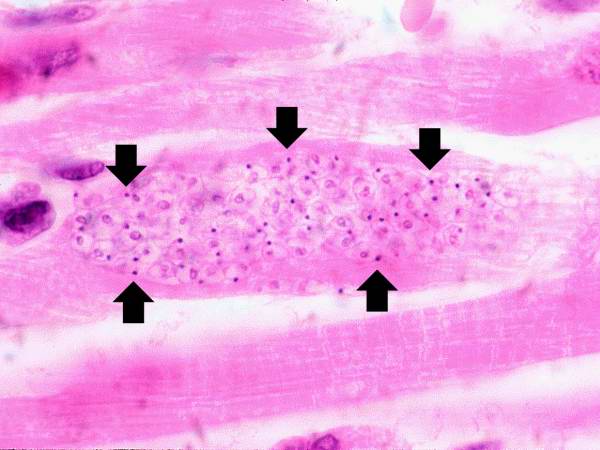
34 KB | This is a higher-power photomicrograph of an H & E stained heart biopsy from this patient. Note the T. cruzi amastigotes (arrows) within this longitudinal section of a myocyte. | 1 |
| 18:05, 19 August 2013 | IPLab6RA1.jpg (file) |  |
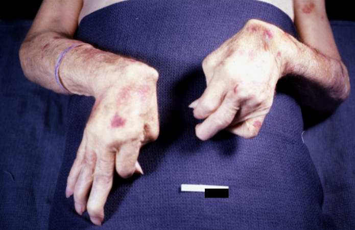
34 KB | This is a gross photograph of the patient's hands at autopsy. Note the swollen joints and the deforming arthritis. | 1 |
| 21:36, 20 August 2013 | IPLab6Amyloid9.jpg (file) |  |
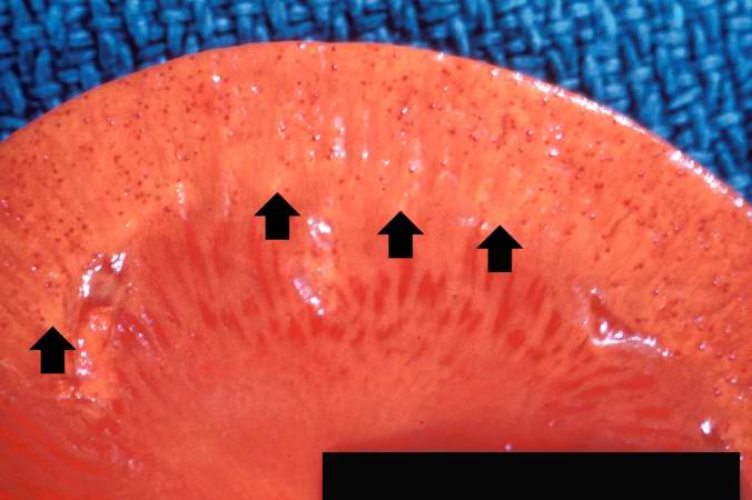
34 KB | This is a gross photograph of kidney from this case. Note the pale yellow material within the cortex (arrows). This is indicative of amyloid within the cortex and the glomeruli. Also note that there are multiple red spots in the cortex. These represent... | 1 |
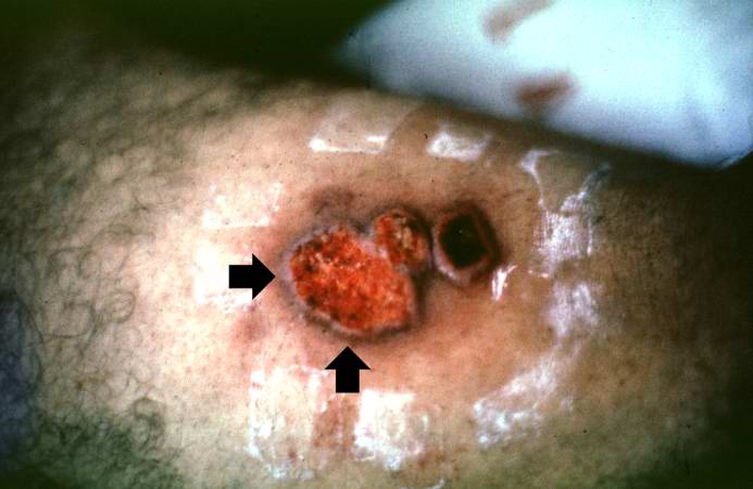
| 04:57, 21 August 2013 | IPLab11Leishmaniasis1.jpg (file) |  |
34 KB | In this photograph of the skin lesion seen in this patient, note the raised edges (arrows) and the ulcerated center. | 1 |
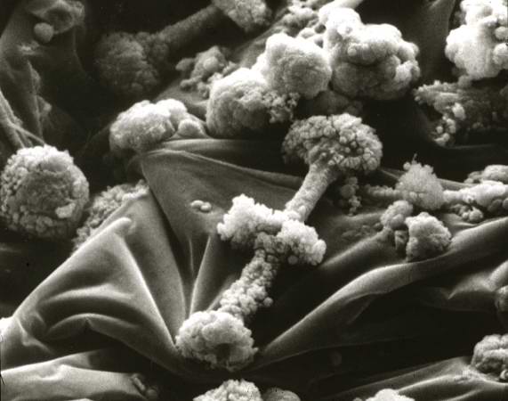
| 05:34, 21 August 2013 | IPLab12Mesothelioma11.jpg (file) |  |
35 KB | Scanning electron micrograph of asbestos bodies. Note the rough surface and the beaded appearance caused by the material adhering to the surface of the asbestos fiber. | 1 |
| 02:50, 21 August 2013 | IPLab8HBV10.jpg (file) |  |
35 KB | This high-power photomicrograph of the previous section shows the HBcAg positive nuclei (arrows). | 1 |
| 03:42, 16 August 2013 | IPLab1Prostate5.jpg (file) |  |
35 KB | This photomicrograph of prostatic epithelium demonstrates an in situ immunohistochemical technique that is used to identify the DNA fragments characteristic of apoptotic nuclei. This technique, terminal deoxynucleotidyl transferase-mediated dUTP-biotin... | 1 |
| 03:39, 19 August 2013 | IPLab3Tuberculosis5.jpg (file) |  |
35 KB | This high-power photomicrograph of a tuberculosis granuloma demonstrates acid-fast bacilli (arrows). | 1 |
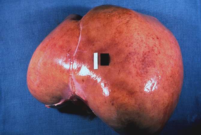
| 21:33, 20 August 2013 | IPLab6Amyloid1.jpg (file) |  |
35 KB | This is a gross picture of liver from this case. Note the pale, swollen appearance of this liver. | 1 |