File list
This special page shows all uploaded files.
| Date | Name | Thumbnail | Size | Description | Versions |
|---|---|---|---|---|---|
| 05:45, 21 August 2013 | IPLab13Myelomeningocele7.jpg (file) |  |
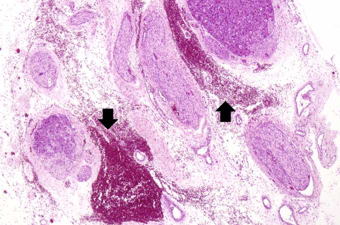
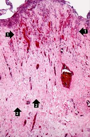
80 KB | This high-power photomicrograph of the spinal cord within the vertebral column shows the hemorrhage (arrows) in this region. | 1 |
| 05:45, 21 August 2013 | IPLab13Myelomeningocele6.jpg (file) |  |
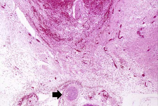
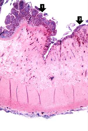
77 KB | This is a high-power photomicrograph of the spinal cord (arrow) immediately beneath the area of hemorrhage. | 1 |
| 05:45, 21 August 2013 | IPLab13Myelomeningocele5.jpg (file) |  |
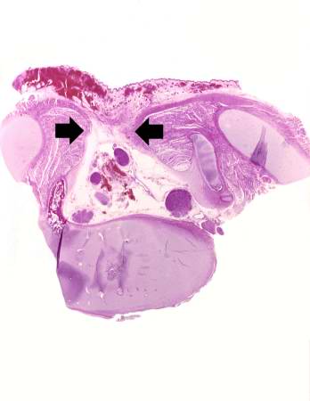
49 KB | This is a higher-power photomicrograph of one of the vertebral bodies from this case. The defect (arrows) in the vertebral body is seen more clearly. The spinal cord is disrupted and there are areas of hemorrhage in this region. | 1 |
| 05:45, 21 August 2013 | IPLab13Myelomeningocele4.jpg (file) |  |
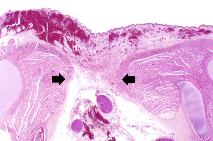
16 KB | This is a low-power photomicrograph of one of the vertebral bodies from this case. In this section there are defects (arrows) in the vertebral body but the skin can be seen over the open vertebral canal. | 1 |
| 05:45, 21 August 2013 | IPLab13Myelomeningocele3.jpg (file) |  |
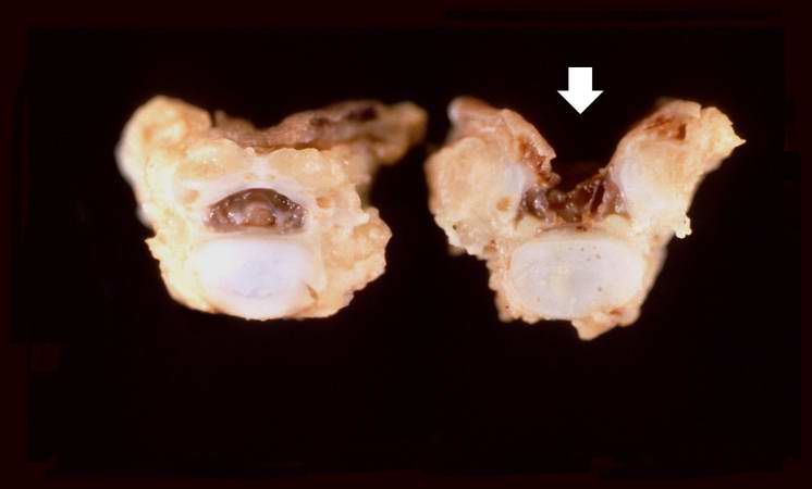
17 KB | This is a closer view of the previous gross photograph showing a normal lumbar vertebra from this case on the left. Once again, note the defect (arrow) in the vertebral body on the right due to failure of the vertebral column to close properly. | 1 |
| 05:44, 21 August 2013 | IPLab13Myelomeningocele2.jpg (file) |  |
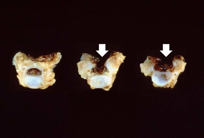
15 KB | This gross photograph shows consecutive lumbar vertebra from this case. Note the defect (arrows) in the two vertebral bodies on the right. This defect was caused by failure of the vertebral column to properly close. | 1 |
| 05:44, 21 August 2013 | IPLab13Myelomeningocele1.jpg (file) |  |
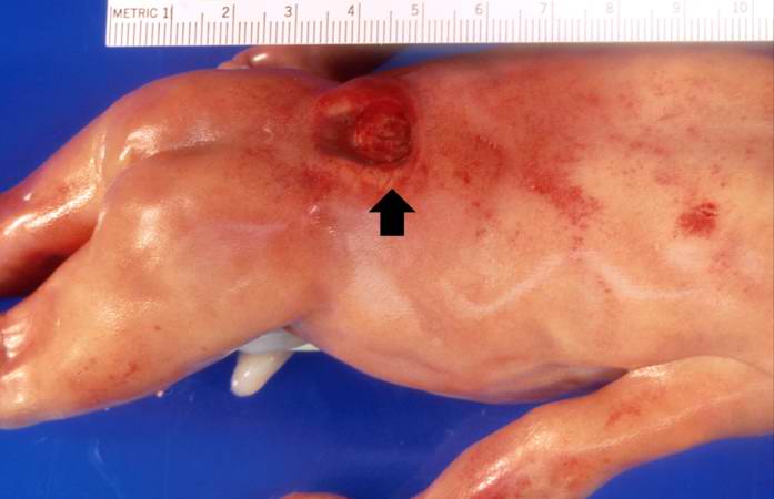
27 KB | This is a gross photograph of the fetus at autopsy. Note the defect in the lower lumbar region of the spinal column (arrow). The myelomeningocele can be seen protruding from this defect. | 1 |
| 05:42, 21 August 2013 | IPLab12COPD5.jpg (file) |  |
50 KB | This gross photograph of the heart taken at autopsy demonstrates right ventricular hypertrophy and dilatation (arrows). | 1 |
| 05:42, 21 August 2013 | IPLab12COPD4.jpg (file) |  |
68 KB | This higher-power photomicrograph of the lung shows more clearly the enlarged air spaces indicative of emphysematous change. | 1 |
| 05:42, 21 August 2013 | IPLab12COPD3.jpg (file) |  |
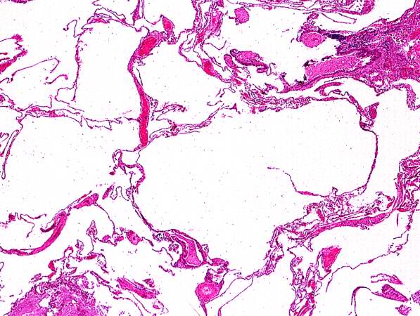
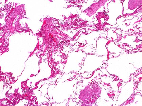
73 KB | This low-power photomicrograph of the lung demonstrates the enlarged air spaces indicative of emphysematous change. | 1 |
| 05:42, 21 August 2013 | IPLab12COPD2.jpg (file) |  |
70 KB | This is a higher-power view of the lung showing the emphysematous change. | 1 |
| 05:41, 21 August 2013 | IPLab12COPD1.jpg (file) |  |
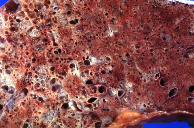
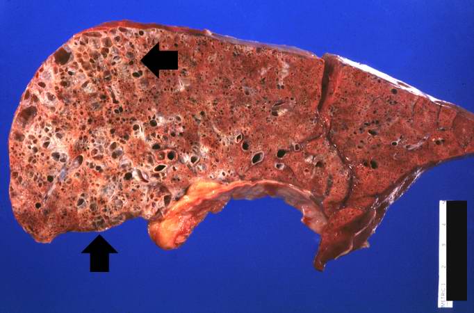
45 KB | This gross photograph of lung taken at autopsy demonstrates the degree of emphysematous change (arrows). Also note that the rest of the lung is consolidated, indicating a pneumonia. | 1 |
| 05:40, 21 August 2013 | IPLab12Burns13.jpg (file) |  |
66 KB | 1 | |
| 05:39, 21 August 2013 | IPLab12Burns12.jpg (file) |  |
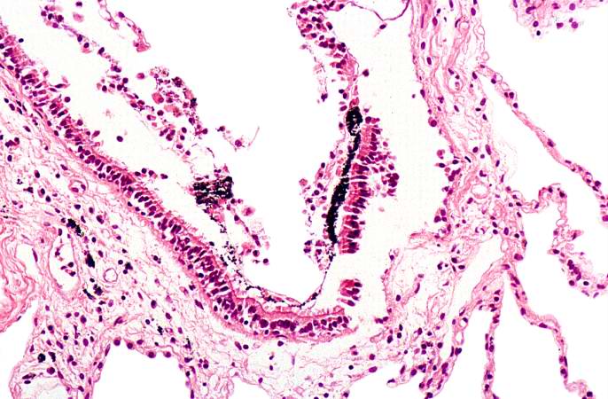
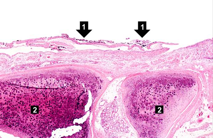
69 KB | This photomicrograph of the trachea shows sloughing of the tracheal epithelium and the black carbonaceous material contained therein (1). This degree of tracheal epithelial damage is indicative of severe inhalation injury. The tracheal cartilage rings ... | 1 |
| 05:39, 21 August 2013 | IPLab12Burns11.jpg (file) |  |
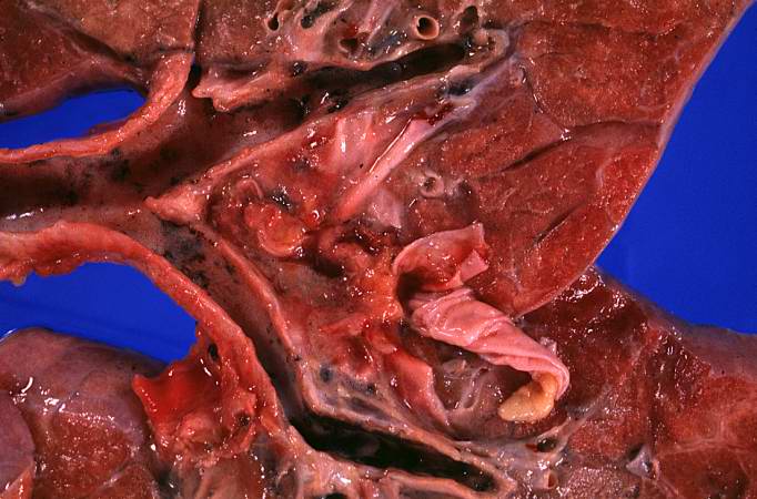
56 KB | This closer view of the lung shows the black carbonaceous material in the trachea as well as in the main stem bronchi. | 1 |
| 05:39, 21 August 2013 | IPLab12Burns10.jpg (file) |  |
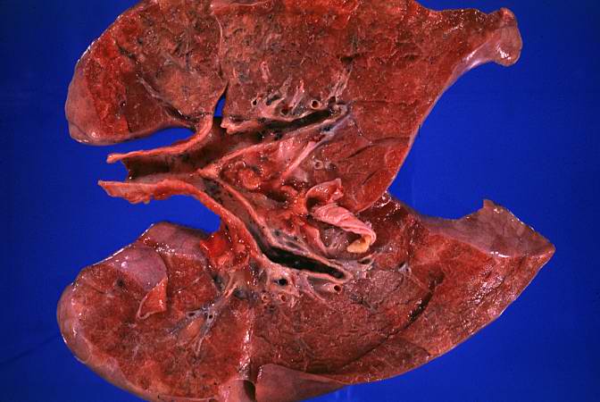
45 KB | This photograph of the lung again shows the black carbonaceous material in the trachea as well as in the main stem bronchi. The lungs are mildly congested and hyperemic. The patient lived for less than 8 hours after the burn injury so there was not eno... | 1 |
| 05:39, 21 August 2013 | IPLab12Burns9.jpg (file) |  |
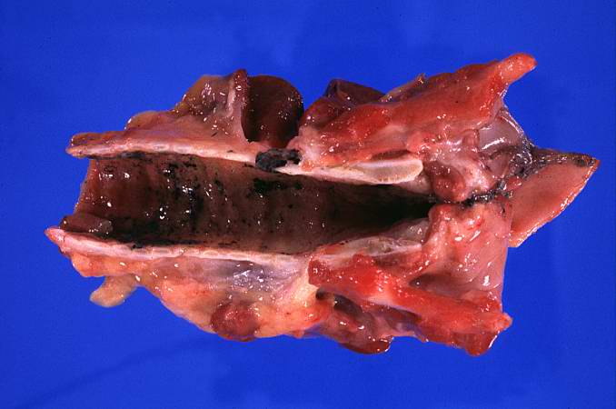
34 KB | This photograph demonstrates the black carbonaceous material in the trachea. | 1 |
| 05:38, 21 August 2013 | IPLab12Burns8.jpg (file) |  |
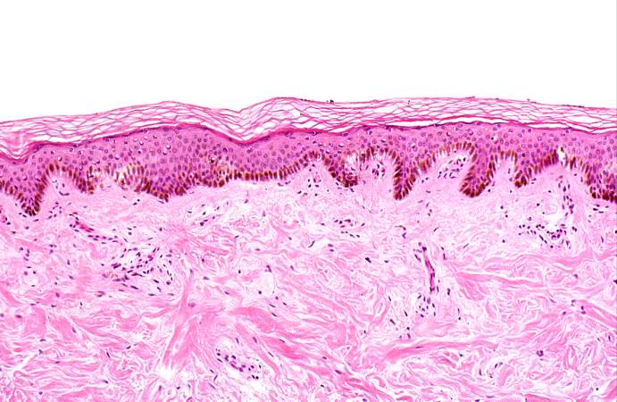
61 KB | This medium-power photomicrograph of the burned skin shows the mild damage to the superficial layers of the epidermis. | 1 |
| 05:38, 21 August 2013 | IPLab12Burns7.jpg (file) |  |
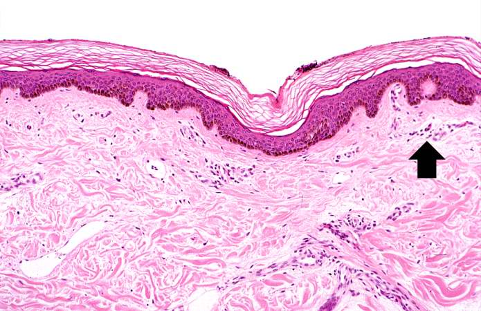
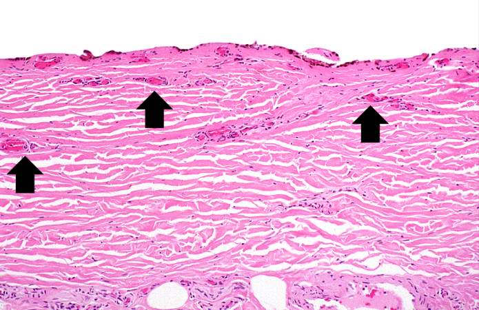
68 KB | This photomicrograph of the burned skin depicts an area of first degree burn. Note that there are no blisters and no damage to the dermis. There is mild damage to the superficial epidermis and some hyperemia (arrow). | 1 |
| 05:38, 21 August 2013 | IPLab12Burns6.jpg (file) |  |
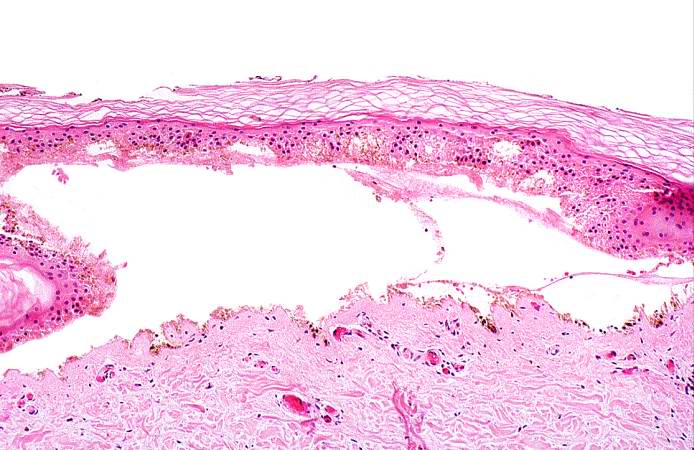
63 KB | This high-power photomicrograph of the burned skin shows the blister and the thrombosed vessels in the dermis. | 1 |
| 05:38, 21 August 2013 | IPLab12Burns5.jpg (file) |  |
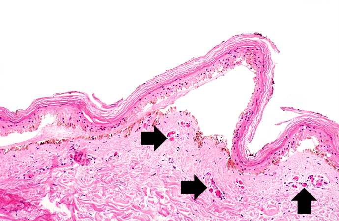
56 KB | This medium-power photomicrograph of the burned skin demonstrates blister formation. The vessels in the dermis are congested (arrows). | 1 |
| 05:37, 21 August 2013 | IPLab12Burns4.jpg (file) |  |
68 KB | This high-power photomicrograph shows the denuded surface of the skin with thrombosed blood vessels (arrows) and necrosis of the dermis. | 1 |
| 05:37, 21 August 2013 | IPLab12Burns3.jpg (file) |  |
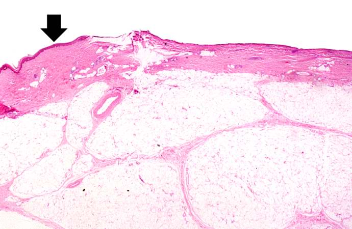
44 KB | This photomicrograph of the burned skin depicts an area of third degree burn. There is some residual epidermis (arrow) but in the majority of this section the epidermis is gone. Also note the severe subcutaneous edema. | 1 |
| 05:37, 21 August 2013 | IPLab12Burns2.jpg (file) |  |
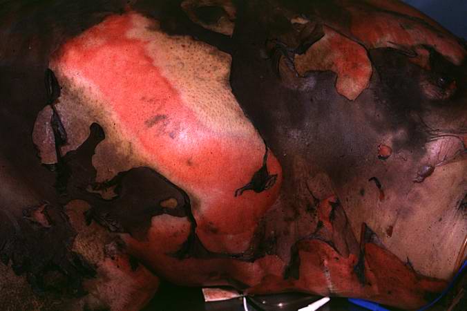
34 KB | This closer view shows the burned skin peeling off to expose the underlying tissue. The various depths of the burn can be appreciated by the color and character of the underlying tissue. | 1 |
| 05:37, 21 August 2013 | IPLab12Burns1.jpg (file) |  |
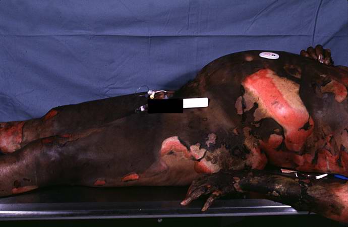
30 KB | This photograph taken at autopsy demonstrates the severity of the surface burns on this patient. | 1 |
| 05:34, 21 August 2013 | IPLab12Mesothelioma11.jpg (file) |  |
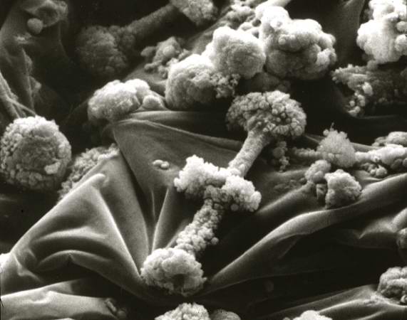
35 KB | Scanning electron micrograph of asbestos bodies. Note the rough surface and the beaded appearance caused by the material adhering to the surface of the asbestos fiber. | 1 |
| 05:34, 21 August 2013 | IPLab12Mesothelioma10.jpg (file) |  |
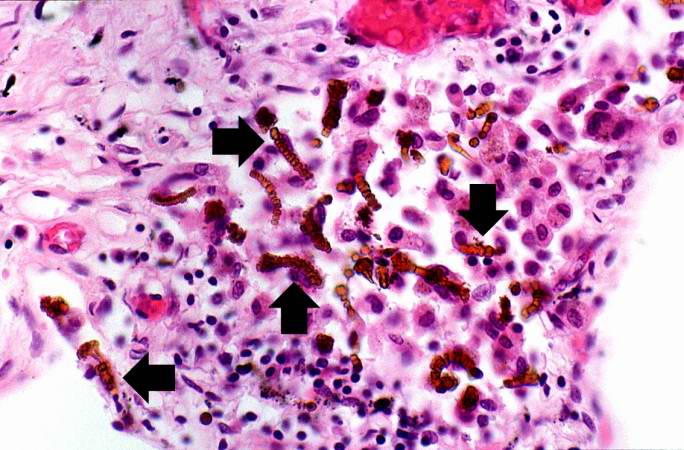
81 KB | Another high-power photomicrograph of the brown asbestos bodies showing the characteristic beaded appearance (arrows). Tumor cells are evident adjacent to the asbestos bodies. | 1 |
| 05:34, 21 August 2013 | IPLab12Mesothelioma9.jpg (file) |  |
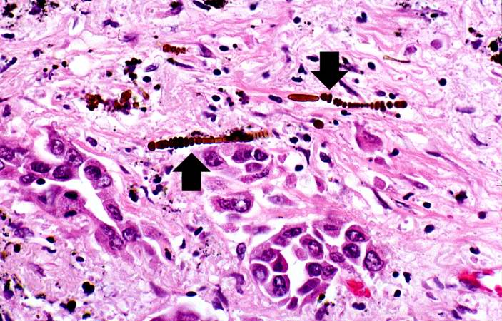
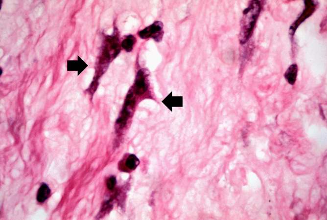
64 KB | This is a high-power photomicrograph of the brown asbestos bodies showing the characteristic beaded appearance (arrows). | 1 |
| 05:33, 21 August 2013 | IPLab12Mesothelioma8.jpg (file) |  |
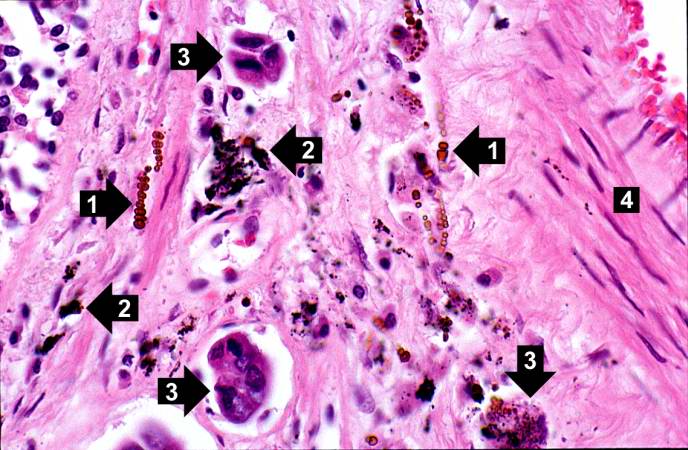
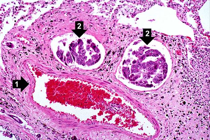
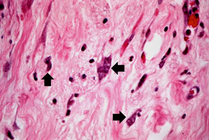
68 KB | This high-power photomicrograph shows brown asbestos bodies (1), accumulations of anthracotic pigment (2), and tumor cells (3) all adjacent to a blood vessel (4). | 1 |
| 05:33, 21 August 2013 | IPLab12Mesothelioma7.jpg (file) |  |
100 KB | In this higher-power photomicrograph there is a blood vessel (1) and adjacent lymphatics that contain tumor cells (2). There are also accumulations of brown material adjacent to these vessels. | 1 |
| 05:33, 21 August 2013 | IPLab12Mesothelioma6.jpg (file) |  |
92 KB | In this higher-power photomicrograph the clusters of tumor cells (arrows) can be appreciated. | 1 |
| 05:33, 21 August 2013 | IPLab12Mesothelioma5.jpg (file) |  |
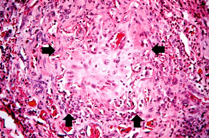
99 KB | This higher-power photomicrograph of left lung shows areas of consolidation and fibrosis (arrows). Also note that in many of these areas there are clusters of blue cells. | 1 |
| 05:33, 21 August 2013 | IPLab12Mesothelioma4.jpg (file) |  |
113 KB | This is a low-power photomicrograph of a section of the left lung. At this magnification you can see areas of consolidation (arrows), especially around blood vessels. | 1 |
| 05:32, 21 August 2013 | IPLab12Mesothelioma3.jpg (file) |  |
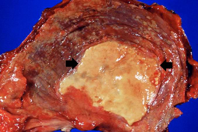
51 KB | This is a gross photograph of the thoracic surface of the diaphragm demonstrating the distinctive fibrocalcific plaque (arrows) produced by mesothelioma and multiple smaller plaques over the pleural surface of the diaphragm. | 1 |
| 05:32, 21 August 2013 | IPLab12Mesothelioma2.jpg (file) |  |
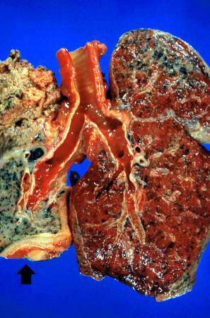
30 KB | This is a gross photograph of cut sections of the lungs. The right lung is congested. The left lung is shrunken and filled with tumor. There is a thick layer of tumor along the inferior portion of the lung (arrow). | 1 |
| 05:32, 21 August 2013 | IPLab12Mesothelioma1.jpg (file) |  |
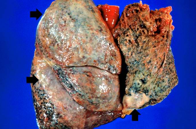
52 KB | This is a gross photograph of the lungs removed at autopsy. There is thickening of the pleural surface due to the tumor (arrows). The apical portion of the left lobe of the lung was ripped while trying to sever the adhesions between the lung and the ch... | 1 |
| 05:30, 21 August 2013 | IPLab12RadiationChanges8.jpg (file) |  |
38 KB | This high-power photomicrograph of the wall of the ileum shows more examples of pleomorphic cells caused by radiation injury (arrows). | 1 |
| 05:29, 21 August 2013 | IPLab12RadiationChanges7.jpg (file) |  |
47 KB | This high-power photomicrograph of the wall of the ileum shows more examples of pleomorphic cells (arrows) due to radiation injury. | 1 |
| 05:29, 21 August 2013 | IPLab12RadiationChanges5.jpg (file) |  |
81 KB | This is a high-power photomicrograph of the wall of the ileum showing a blood vessel that has suffered radiation-induced damage and is completely occluded (arrows). | 1 |
| 05:28, 21 August 2013 | IPLab12RadiationChanges6.jpg (file) |  |
57 KB | This is a high-power photomicrograph of the wall of the ileum showing a blood vessel that has suffered radiation-induced damage and is completely occluded (arrows). | 1 |
| 05:28, 21 August 2013 | IPLab12RadiationChanges4.jpg (file) |  |
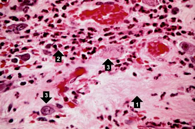
39 KB | This is a high-power photomicrograph showing the atrophied epithelium in the area of radiation injury (1). Note the dense fibrous connective tissue within the wall of the ileum and the congested blood vessels (2). | 1 |
| 05:28, 21 August 2013 | IPLab12RadiationChanges3.jpg (file) |  |
38 KB | This is a higher-power photomicrograph showing the atrophied epithelium in the area of radiation injury. There are some epithelial cells deep within the mucosa (1). Note the dense fibrous connective tissue (2) within the wall of the ileum. | 1 |
| 05:28, 21 August 2013 | IPLab12RadiationChanges2.jpg (file) |  |
26 KB | This is a higher-power photomicrograph of the surgical specimen of the ileum showing the transition from the normal epithelium (1) to the atrophied epithelium (2) in the area of radiation injury. | 1 |
| 05:27, 21 August 2013 | IPLab12RadiationChanges1.jpg (file) |  |
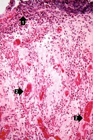
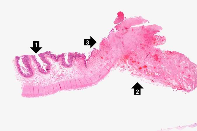
26 KB | This is a low-power photomicrograph of the surgical specimen of the ileum. The normal ileum is to the left (1). The area of stricture consists of dense fibrous connective tissue (2) and there is loss or marked atrophy of the epithelium (3). | 1 |
| 05:25, 21 August 2013 | IPLab12RadiationFibrosis13.jpg (file) |  |
69 KB | This is a high-power photomicrograph of a recanalized blood vessel in the lung. Notice the anthracotic pigment adjacent to the vessel (arrows). | 1 |
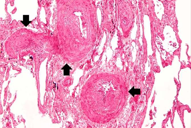
| 05:25, 21 August 2013 | IPLab12RadiationFibrosis12.jpg (file) |  |
72 KB | This high-power photomicrograph shows intimal changes (arrows) in this blood vessel in the lung. | 1 |
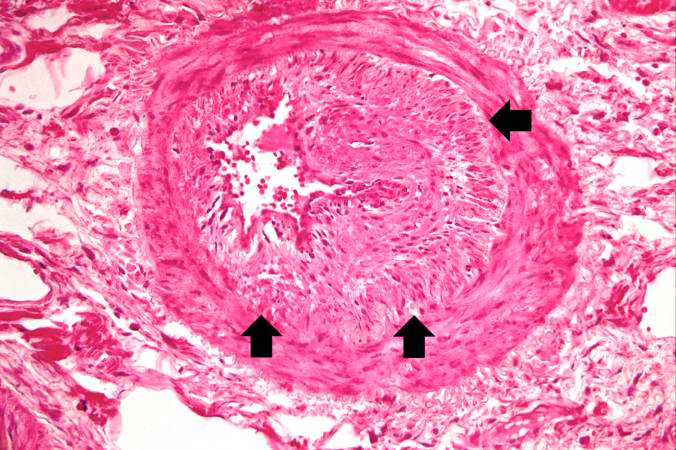
| 05:25, 21 August 2013 | IPLab12RadiationFibrosis11.jpg (file) |  |
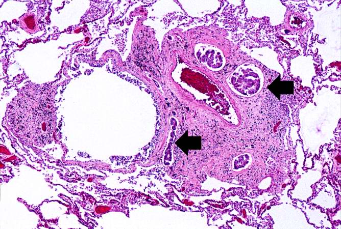
73 KB | This medium-power photomicrograph shows fibrosis and severe intimal changes in blood vessels (arrows). | 1 |
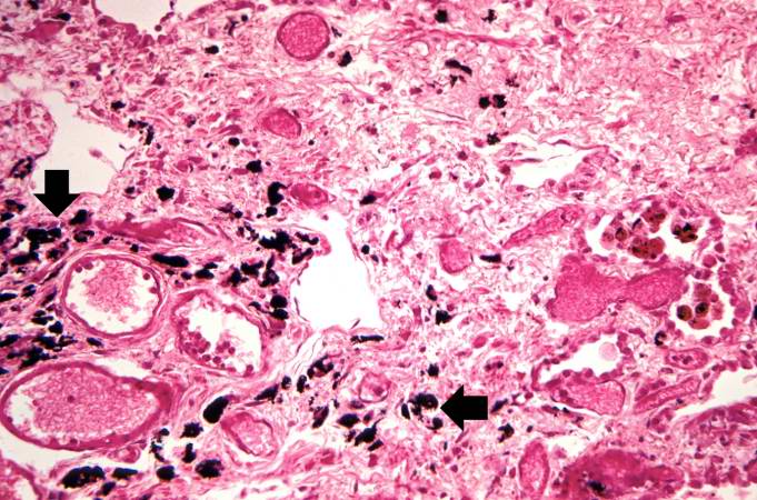
| 05:25, 21 August 2013 | IPLab12RadiationFibrosis10.jpg (file) |  |
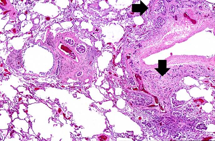
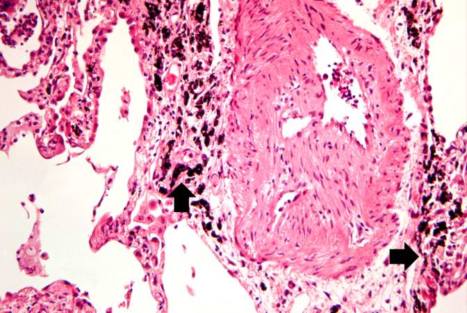
80 KB | This is another high-power photomicrograph of an area of tissue with diffuse fibrosis and thickening of the alveolar septa. There are also accumulations of anthracotic pigment in this area (arrows). | 1 |
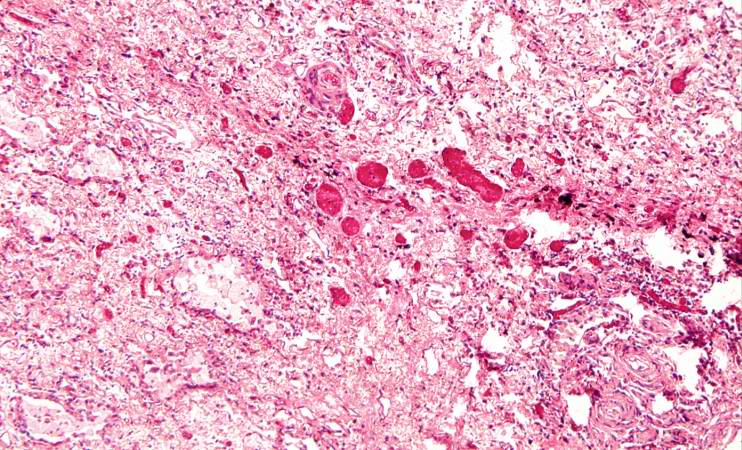
| 05:25, 21 August 2013 | IPLab12RadiationFibrosis9.jpg (file) |  |
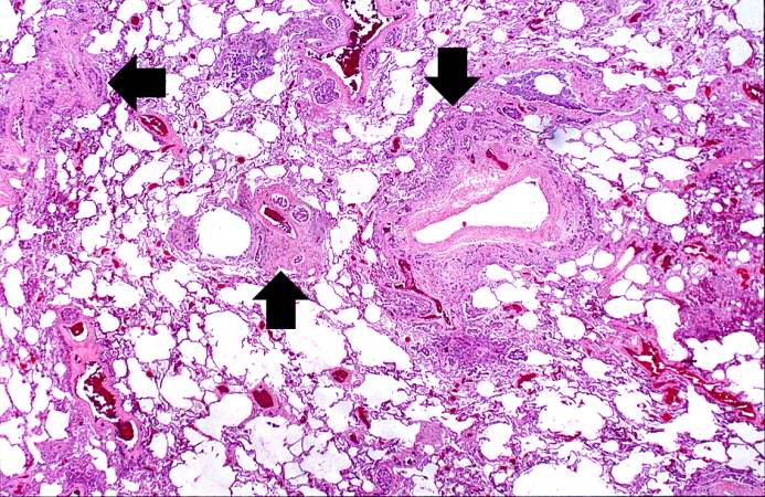
107 KB | This is a photomicrograph of an area of tissue exhibiting diffuse fibrosis and thickening of the alveolar septa. | 1 |
| 05:24, 21 August 2013 | IPLab12RadiationFibrosis8.jpg (file) |  |
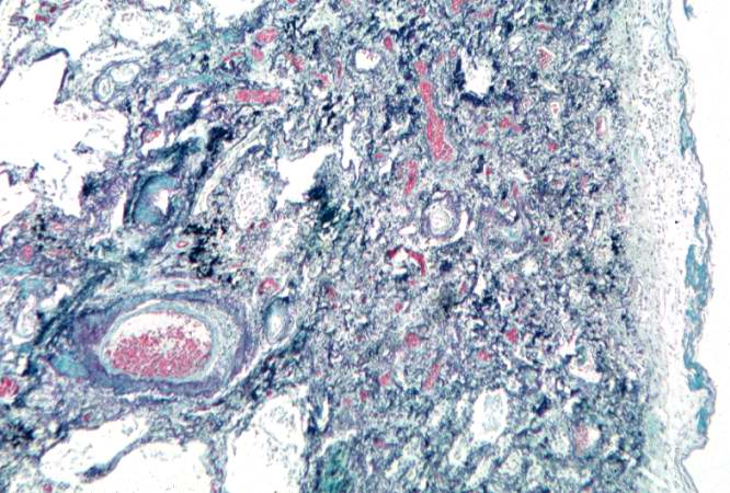
75 KB | This is a photomicrograph of a trichrome-stained section of lung demonstrating the extensive fibrosis throughout this section (green-blue stained material is fibrous connective tissue). | 1 |