File list
This special page shows all uploaded files.
| Date | Name | Thumbnail | Size | Description | Versions |
|---|---|---|---|---|---|
| 15:20, 20 August 2013 | IPLab5Gout6.jpg (file) |  |
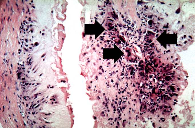
63 KB | This is a high-power photomicrograph of the edge of the tophus. The character of the intense chronic inflammatory cell reaction is evident and note the presence of giant cells within this inflammatory cell reaction (arrows). | 1 |
| 15:20, 20 August 2013 | IPLab5Gout7.jpg (file) |  |
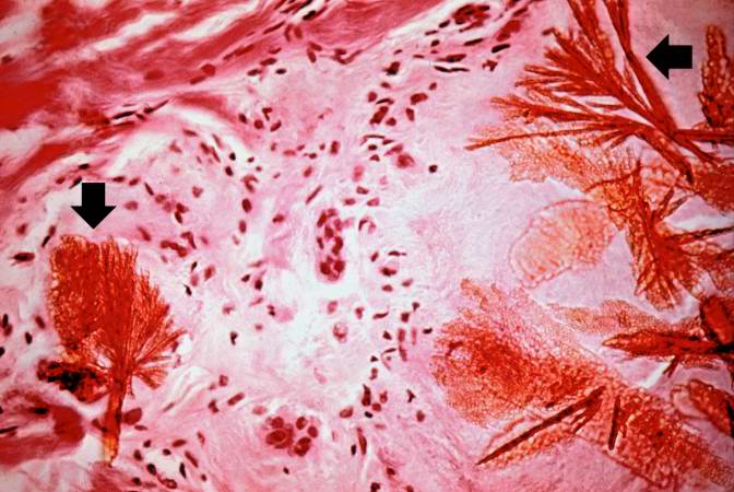
59 KB | This is a photomicrograph of a tophus that was fixed in alcohol prior to histologic processing. The alcohol fixation preserves the water soluble urate crystals within the tissue. Note the urate crystals visible in this photomicrograph (arrows). Also no... | 1 |
| 15:20, 20 August 2013 | IPLab5Gout8.jpg (file) |  |
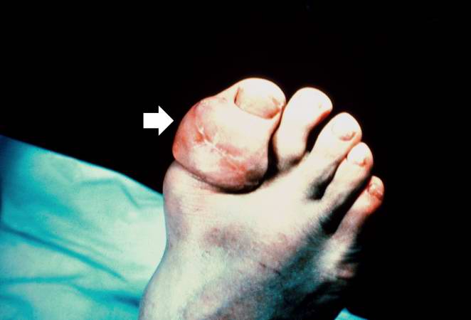
20 KB | This is a gross photograph of a tophus on the great toe of another patient with gout (arrow). The healed surgical incision and the size of this tophus indicate that this was a long-standing problem for this patient. | 1 |
| 15:26, 20 August 2013 | IPLab5DM1.jpg (file) |  |
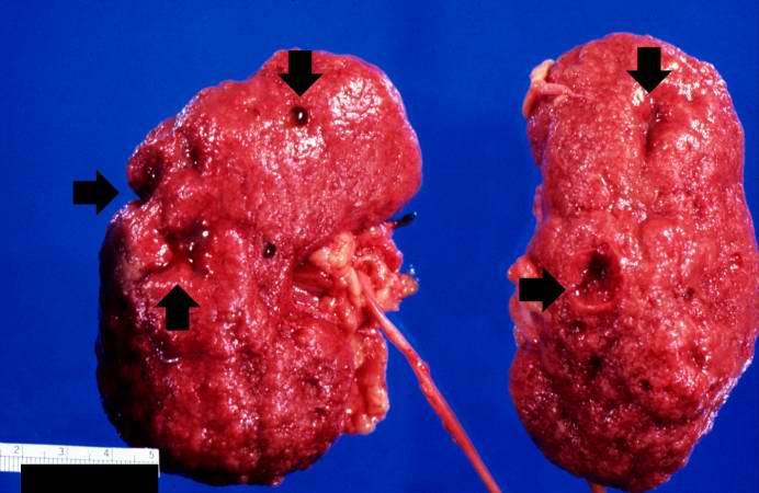
44 KB | This is a gross photograph of the kidneys from this case. Note that there are multiple shrunken regions (old infarcts) (arrows) and the kidneys have a rough granular appearance on the surface, which is caused by multiple small infarcts of small vessels... | 1 |
| 15:26, 20 August 2013 | IPLab5DM2.jpg (file) |  |
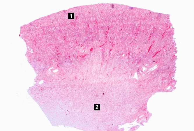
36 KB | This is a low-power photomicrograph of the kidney from this patient. The section extends from cortex (1) to the medulla (2). | 1 |
| 15:27, 20 August 2013 | IPLab5DM3.jpg (file) |  |
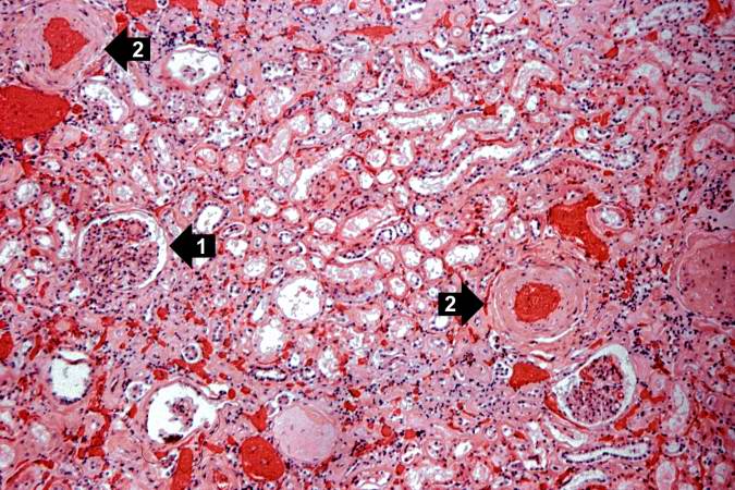
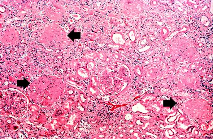
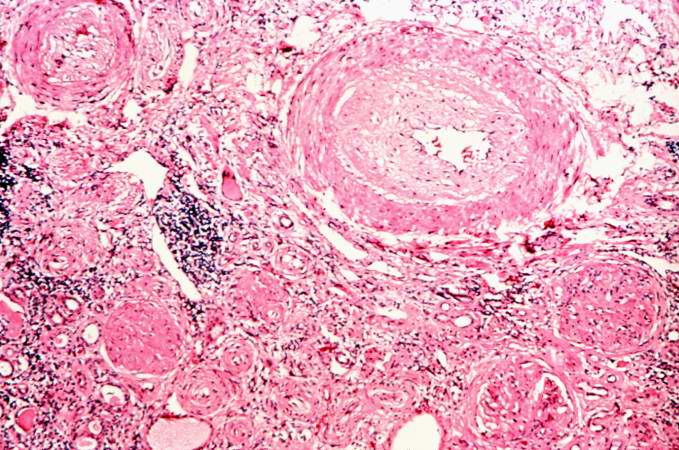
94 KB | This is a higher-power photomicrograph of the cortical region. In this region there is ischemic obsolescence of glomeruli and one glomerulus with nodular glomerulosclerosis (1). Also note the thickened walls of the blood vessels (2). | 1 |
| 15:27, 20 August 2013 | IPLab5DM4.jpg (file) |  |
74 KB | This is a high-power photomicrograph of two glomeruli with intercapillary glomerulosclerosis (arrows). | 1 |
| 15:27, 20 August 2013 | IPLab5DM5.jpg (file) |  |
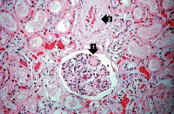
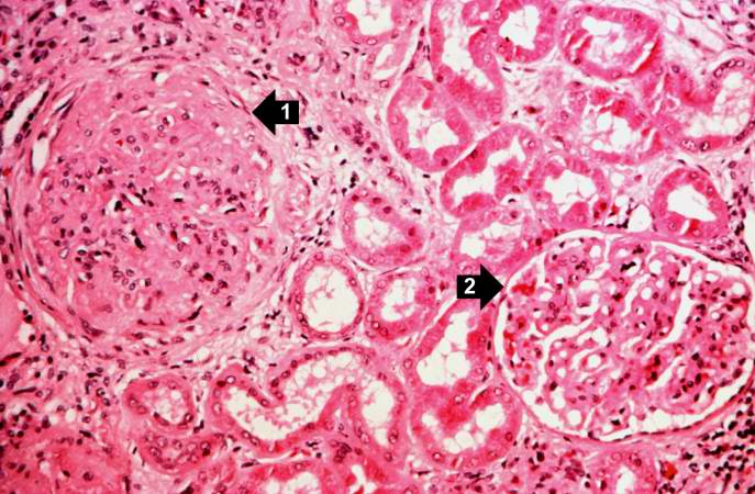
74 KB | This is a photomicrograph of a glomerulus with nodular glomerulosclerosis (1). Also note the intertubular fibrosis and the changes in the blood vessels (2). | 1 |
| 15:28, 20 August 2013 | IPLab5DM6.jpg (file) |  |
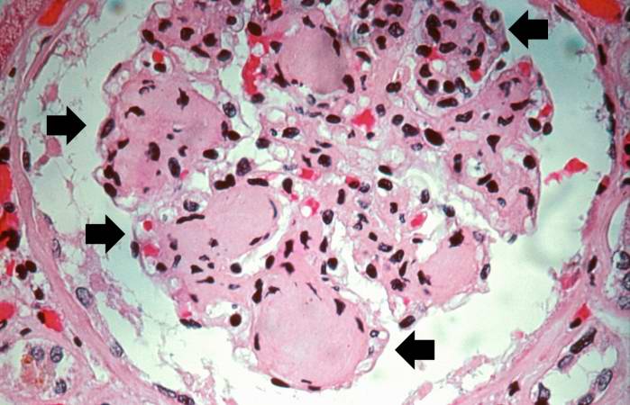
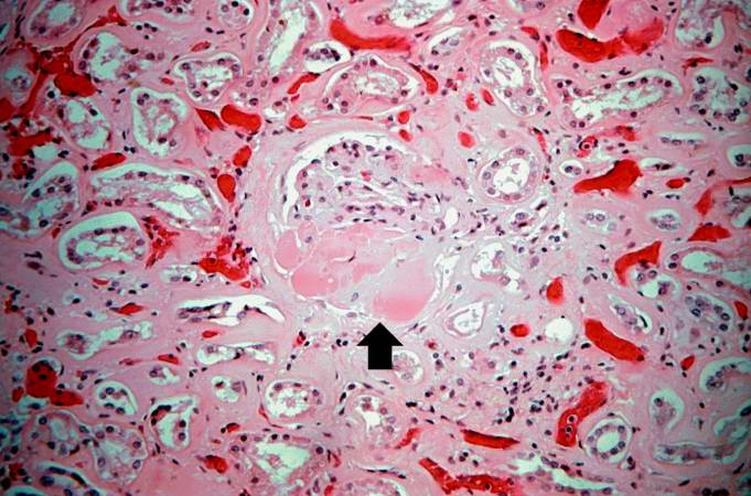
56 KB | This is a higher-power photomicrograph of a glomerulus with nodular glomerulosclerosis (arrows). These are the classic Kimmelstiel-Wilson lesions ("K-W lesions") seen in diabetics with nodular glomerulosclerosis. | 1 |
| 15:29, 20 August 2013 | IPLab5DM7.jpg (file) |  |
66 KB | This is a photomicrograph of kidney with a focal exudative lesion in a glomerulus (arrow) and sclerosis, interstitial fibrosis, and congestion. | 1 |
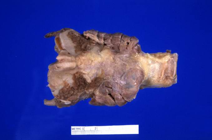
| 17:42, 20 August 2013 | IPLab6Hashimoto1.jpg (file) | 20 KB | This is a gross photograph of thyroid gland taken at autopsy. The gland is only slightly enlarged and has a firm texture. | 1 | |
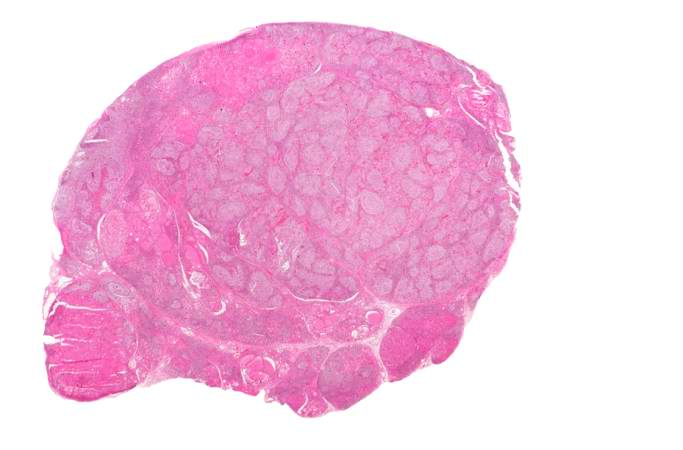
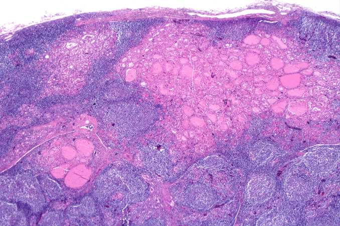
| 17:42, 20 August 2013 | IPLab6Hashimoto2.jpg (file) | 27 KB | This is a low-power photomicrograph of thyroid from this case. Note that the tissue is more cellular than one would expect and there does not appear to be normal colloid-filled blue spaces in this gland. | 1 | |
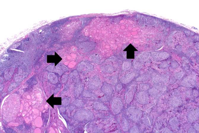
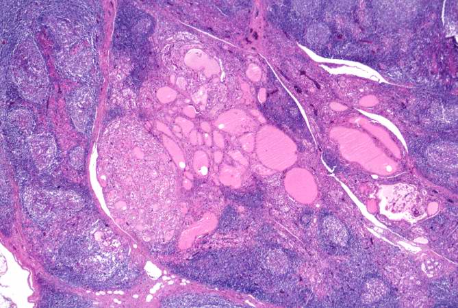
| 17:43, 20 August 2013 | IPLab6Hashimoto3.jpg (file) | 55 KB | This is a higher-power photomicrograph of thyroid from this case. Note the large number of blue-staining inflammatory cells in this tissue. These cells appear to be forming germinal centers. Some residual thyroid gland tissue can be seen in this sectio... | 1 | |
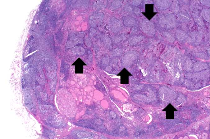
| 17:43, 20 August 2013 | IPLab6Hashimoto4.jpg (file) | 58 KB | This is another view of thyroid gland filled with inflammatory cells forming germinal centers (arrows). | 1 | |
| 17:44, 20 August 2013 | IPLab6Hashimoto5.jpg (file) | 76 KB | This is a higher-power photomicrograph of thyroid from this case showing the inflammatory cells and the residual thyroid tissue. | 1 | |
| 17:46, 20 August 2013 | IPLab6Hashimoto6.jpg (file) | 75 KB | This is another higher-power photomicrograph of thyroid from this case showing the inflammatory cells and the residual thyroid tissue. | 1 | |
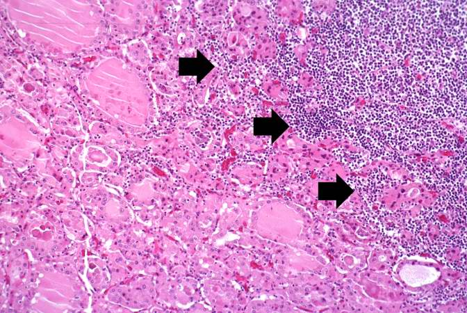
| 17:46, 20 August 2013 | IPLab6Hashimoto7.jpg (file) | 87 KB | This is a high-power photomicrograph showing the inflammatory cells infiltrating into the residual thyroid tissue (arrows). | 1 | |
| 17:47, 20 August 2013 | IPLab6Hashimoto8.jpg (file) | 94 KB | This is a high-power photomicrograph showing the lymphocytes and plasma cells surrounding the thyroid gland epithelium. | 1 | |
| 17:47, 20 August 2013 | IPLab6Hashimoto9.jpg (file) | 111 KB | This high-power photomicrograph shows more clearly the lymphocytes and plasma cells surrounding the thyroid gland epithelium. Large, eosinophilic, degenerating thyroid gland cells (Hurthle cells) can be seen in this section (arrows). | 1 | |
| 17:55, 20 August 2013 | IPLab6PAN1.jpg (file) |  |
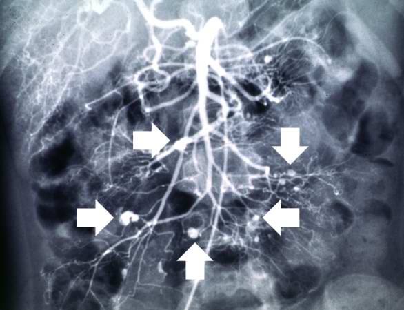
36 KB | This angiogram of the abdominal viscera demonstrates numerous aneurysms throughout the mesenteric circulation (arrows). | 1 |
| 17:55, 20 August 2013 | IPLab6PAN2.jpg (file) |  |
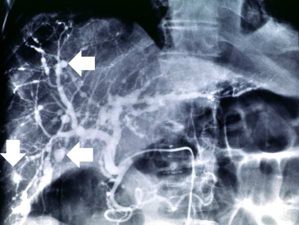
36 KB | This angiogram of the liver also demonstrates numerous aneurysms throughout the hepatic circulation (arrows). | 1 |
| 17:56, 20 August 2013 | IPLab6PAN3.jpg (file) |  |
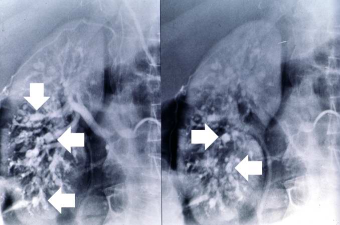
36 KB | This angiogram of the kidneys demonstrates numerous aneurysmal dilatations in the renal circulation (arrows). | 1 |
| 17:56, 20 August 2013 | IPLab6PAN4.jpg (file) |  |
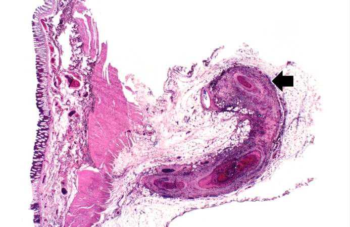
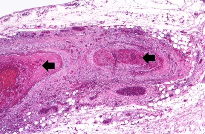
51 KB | This is a low-power photomicrograph of a mesenteric vessel from this case of polyarteritis nodosa (arrow). The vessel is completely occluded by thrombotic material and the vessel wall is infiltrated with inflammatory cells. | 1 |
| 17:56, 20 August 2013 | IPLab6PAN5.jpg (file) |  |
79 KB | This is a higher-power photomicrograph of this mesenteric vessel. Note the thrombotic material occluding the vessel (arrows) and the inflammatory cell infiltrate in the wall of the vessel and in the surrounding adventitia. | 1 |
| 17:57, 20 August 2013 | IPLab6PAN6.jpg (file) |  |
50 KB | his is another example of a mesenteric artery from this case. There is a marked inflammatory cell response surrounding this vessel, fresh hemorrhage (1), and thrombotic material (2). | 1 |
| 17:57, 20 August 2013 | IPLab6PAN7.jpg (file) |  |
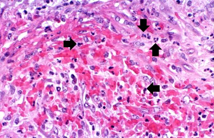
67 KB | This is a high-power photomicrograph of the vessel wall. There is hemorrhage and infiltration with inflammatory cells--primarily neutrophils (arrows). | 1 |
| 17:58, 20 August 2013 | IPLab6PAN8.jpg (file) |  |
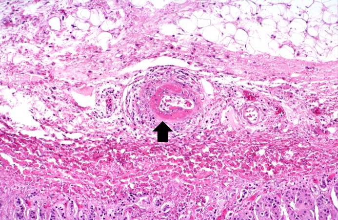
86 KB | This is a high-power photomicrograph of a small vessel with a rim of fibrinoid necrosis (arrow). | 1 |
| 17:58, 20 August 2013 | IPLab6PAN9.jpg (file) |  |
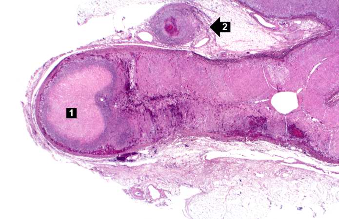
53 KB | This is a low-power photomicrograph of the adrenal gland. There is an area of necrosis in the adrenal (1) and an affected vessel adjacent to the adrenal (2). | 1 |
| 17:59, 20 August 2013 | IPLab6PAN10.jpg (file) |  |
76 KB | This is a higher-power photomicrograph of the affected vessel from the previous image. The vessel wall is infiltrated with inflammatory cells and the vessel lumen is completely occluded (arrow). | 1 |
| 17:59, 20 August 2013 | IPLab6PAN11.jpg (file) |  |
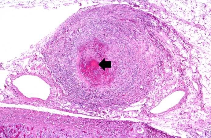
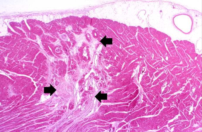
69 KB | This is a low-power photomicrograph of the heart. There are areas of fibrosis in the myocardium (arrows). Note that the large epicardial coronary artery is normal. | 1 |
| 18:00, 20 August 2013 | IPLab6PAN12.jpg (file) |  |
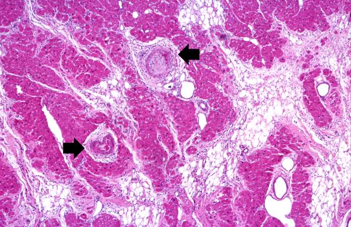
88 KB | This is a higher-power photomicrograph of the affected vessels in the heart (arrows). There are areas of fibrosis (old infarcts) in the myocardium adjacent to these affected vessels. | 1 |
| 18:00, 20 August 2013 | IPLab6PAN13.jpg (file) |  |
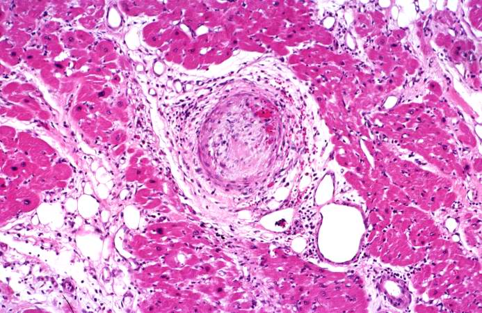
85 KB | This is a high-power photomicrograph of the affected vessel in the heart. The vessel lumen is completely occluded. | 1 |
| 19:57, 20 August 2013 | IPLab6Scleroderma1.jpg (file) |  |
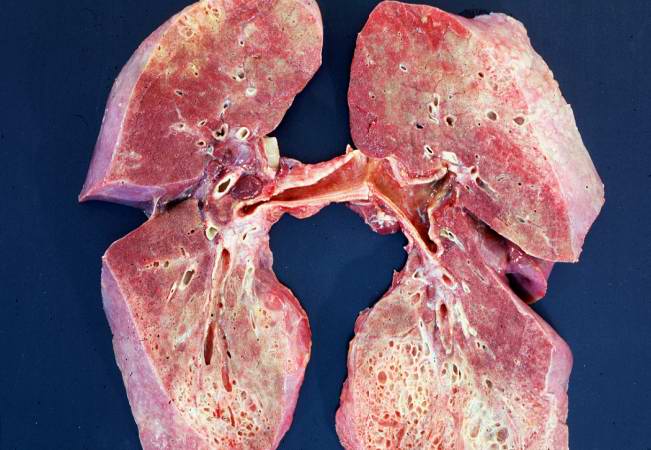
49 KB | This is a gross photograph of cut section of the lungs from this patient. Note the extensive fibrosis of the lung parenchyma. | 1 |
| 19:58, 20 August 2013 | IPLab6Scleroderma2.jpg (file) |  |
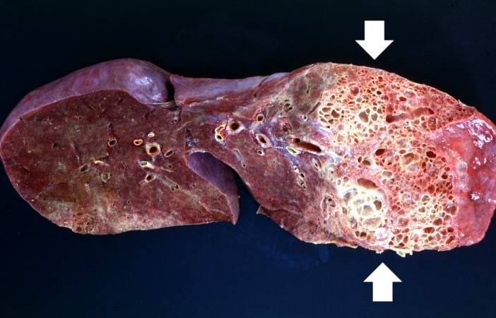
43 KB | This is a gross photograph of a cut section of one lung from this patient. Note the extensive fibrosis lower lobe (arrows). | 1 |
| 19:58, 20 August 2013 | IPLab6Scleroderma3.jpg (file) |  |
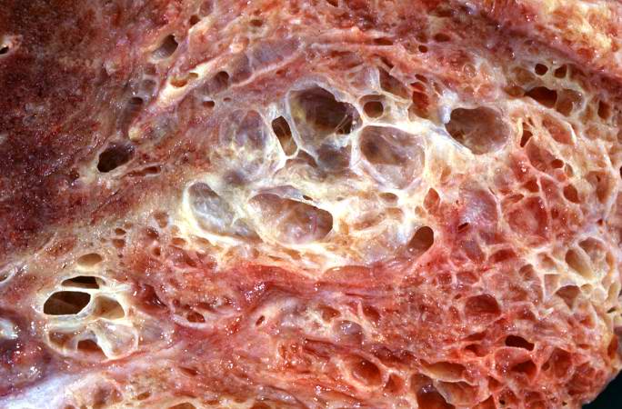
64 KB | This is a closer view of the cut section of lung from this patient. Note the extensive fibrosis and the severe emphysematous changes. | 1 |
| 19:59, 20 August 2013 | IPLab6Scleroderma4.jpg (file) |  |
65 KB | This is a closer view of the cut section of lung from this patient showing the extensive fibrosis and the severe emphysematous change. | 1 |
| 19:59, 20 August 2013 | IPLab6Scleroderma5.jpg (file) |  |
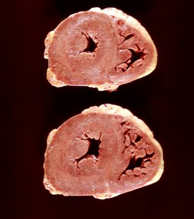
19 KB | This is a gross photograph of the heart from this case. There is thickening of the left ventricular wall and some thickening of the right ventricle as well. | 1 |
| 20:10, 20 August 2013 | IPLab6TB1.jpg (file) |  |
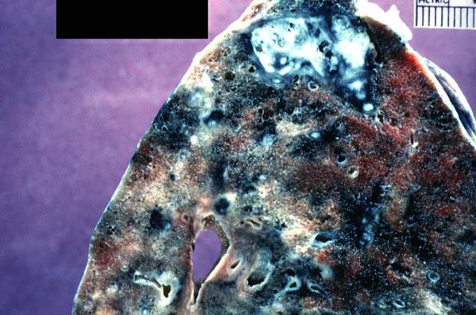
63 KB | This is a photograph of a section of lung with an apical lesion. This lesion represents an old healed lesion from Mycobacterium tuberculosis infection. | 1 |
| 20:10, 20 August 2013 | IPLab6TB2.jpg (file) |  |
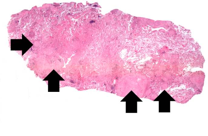
36 KB | This is a low-power photomicrograph of lung tissue with multiple circumscribed nodules - granulomas (arrows). | 1 |
| 20:10, 20 August 2013 | IPLab6TB3.jpg (file) |  |
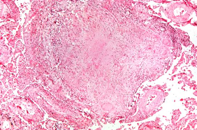
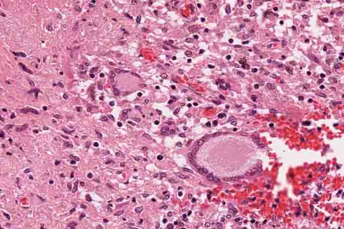
72 KB | This is a higher-power photomicrograph of a TB granuloma. Note the eosinophilic material in the center of this granuloma (caseous necrosis) and the epithelioid macrophages and giant cells around the periphery. | 1 |
| 20:11, 20 August 2013 | IPLab6TB4.jpg (file) |  |
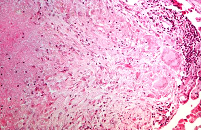
68 KB | This is a higher-power photomicrograph of a TB granuloma. The area of caseous necrosis is on the left side of the image, there are multinucleated giant cells and epithelioid macrophages throughout the remainder of the tissue. | 1 |
| 20:13, 20 August 2013 | IPLab6TB5.jpg (file) |  |
194 KB | High-power photomicrograph of a TB granuloma with multinucleated giant cells adjacent to an area of caseous necrosis (to the left). | 1 |
| 20:14, 20 August 2013 | IPLab6TB6.jpg (file) |  |
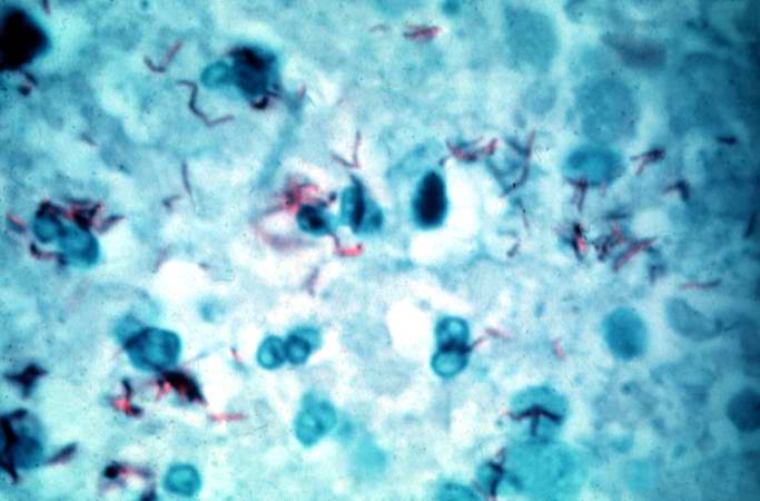
37 KB | This is a high-power (oil immersion) photomicrograph of granuloma stained with an acid-fast stain. Mycobacterium tuberculosis bacilli stain red. | 1 |
| 20:27, 20 August 2013 | IPLab6GN2.jpg (file) |  |
103 KB | This is a higher-power photomicrograph of hyalinized glomeruli (arrows) and glomeruli with thick basement membranes. | 1 |
| 20:27, 20 August 2013 | IPLab6GN3.jpg (file) |  |
71 KB | This is a higher-power photomicrograph of hyalinized glomeruli (1) and glomeruli with thickened basement membranes (2). | 1 |
| 20:28, 20 August 2013 | IPLab6GN4.jpg (file) |  |
87 KB | This is a photomicrograph of interstitial and vascular lesions in end stage renal disease. | 1 |
| 20:28, 20 August 2013 | IPLab6GN5.jpg (file) |  |
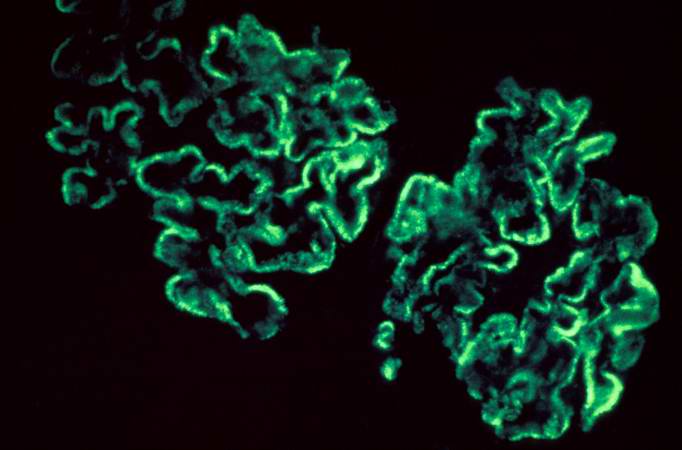
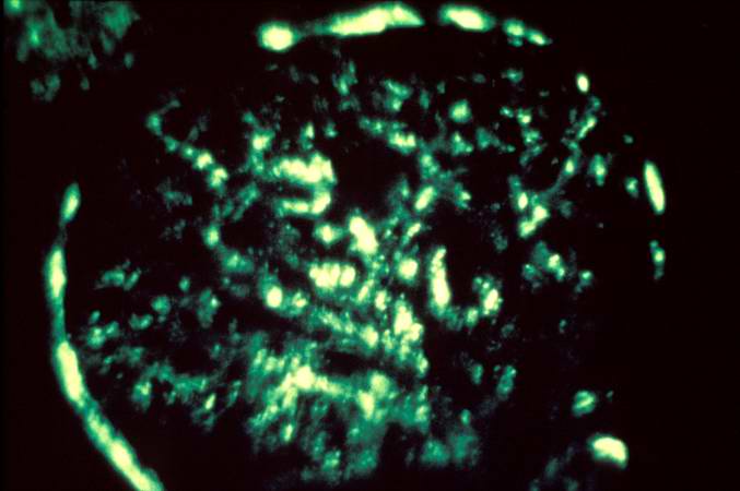
30 KB | This is an immunofluorescent photomicrograph of granular membranous immunofluorescence (immune complex disease). The antibody used for these studies was specific for IgG. | 1 |
| 20:29, 20 August 2013 | IPLab6GN6.jpg (file) |  |
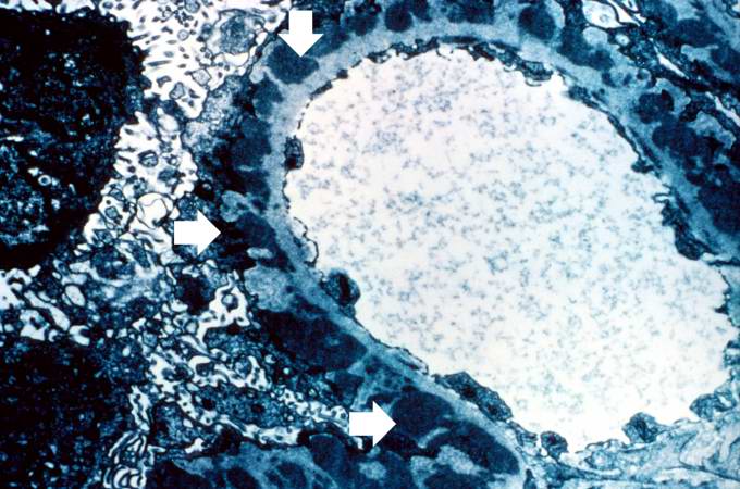
66 KB | This is an electron micrograph of subepithelial granular electron dense deposits (arrows) which correspond to the granular immunofluorescence seen in the previous image. | 1 |
| 20:29, 20 August 2013 | IPLab6GN7.jpg (file) |  |
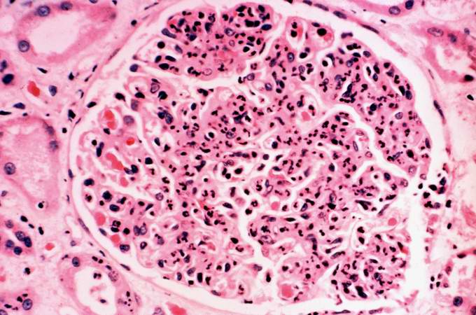
62 KB | This is a photomicrograph of a glomerulus from another case with acute poststreptococcal glomerulonephritis. In this case the immune complex glomerular disease is ongoing with necrosis and accumulation of neutrophils in the glomerulus. | 1 |
| 20:30, 20 August 2013 | IPLab6GN8.jpg (file) |  |
31 KB | This immunofluorescent photomicrograph of a glomerulus from a case of acute poststreptococcal glomerulonephritis shows a granular immunofluorescence pattern consistent with immune complex disease. The primary antibody used for this staining was specifi... | 1 |