Difference between revisions of "IPLab:Lab 4:Atheromatous Emboli"
Seung Park (talk | contribs) (Created page with "== Images == <gallery heights="250px" widths="250px"> File:IPlab4AtheromatousEmboli1.jpg|This is a gross photograph of the aorta from this patient opened lengthwise with the l...") |
Seung Park (talk | contribs) |
||
| Line 1: | Line 1: | ||
== Images == | == Images == | ||
<gallery heights="250px" widths="250px"> | <gallery heights="250px" widths="250px"> | ||
| − | File: | + | File:IPLab4AtheromatousEmboli1.jpg|This is a gross photograph of the aorta from this patient opened lengthwise with the luminal surface visible. Note the rough surface with ulcerations and adherent thrombotic material. There is a mild dilation (aneurysm) at the distal aorta just at the bifurcation with an accumulation of thrombus. |
| − | File: | + | File:IPLab4AtheromatousEmboli2.jpg|This is a closer view of the luminal surface of the aorta from the previous image. The rough, ulcerated surface and the thrombotic material can be easily seen in this image. |
| − | File: | + | File:IPLab4AtheromatousEmboli3.jpg|This is a low-power photomicrograph of kidney tissue. Several blood vessels can be identified at the corticomedullary junction (arrows). |
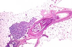
| − | File: | + | File:IPLab4AtheromatousEmboli4.jpg|This higher-power photomicrograph of one of the arcuate arteries reveals a cholesterol embolus. Note the needle-shaped space (arrow) within the lumen of this artery (arrow) which represents the space occupied by the cholesterol crystal that was dissolved away during histologic processing. |
| − | File: | + | File:IPLab4AtheromatousEmboli5.jpg|This is another view of this vessel with an atherosclerotic embolus. Note the cholesterol clefts (1) and thrombotic material (2) that occlude this artery. |
| − | File: | + | File:IPLab4AtheromatousEmboli6.jpg|A mesenteric artery also had an atherosclerotic embolus. Again note the cholesterol clefts and thrombotic material that occlude this artery. |
</gallery> | </gallery> | ||
Revision as of 17:02, 19 August 2013
Images[edit]
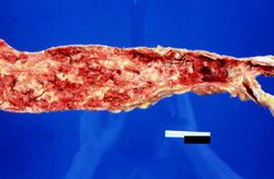
This is a gross photograph of the aorta from this patient opened lengthwise with the luminal surface visible. Note the rough surface with ulcerations and adherent thrombotic material. There is a mild dilation (aneurysm) at the distal aorta just at the bifurcation with an accumulation of thrombus.
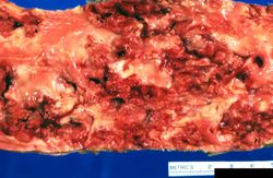
This is a closer view of the luminal surface of the aorta from the previous image. The rough, ulcerated surface and the thrombotic material can be easily seen in this image.
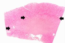
This is a low-power photomicrograph of kidney tissue. Several blood vessels can be identified at the corticomedullary junction (arrows).
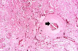
This higher-power photomicrograph of one of the arcuate arteries reveals a cholesterol embolus. Note the needle-shaped space (arrow) within the lumen of this artery (arrow) which represents the space occupied by the cholesterol crystal that was dissolved away during histologic processing.
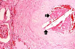
This is another view of this vessel with an atherosclerotic embolus. Note the cholesterol clefts (1) and thrombotic material (2) that occlude this artery.
A mesenteric artery also had an atherosclerotic embolus. Again note the cholesterol clefts and thrombotic material that occlude this artery.


| |||||
A thrombus is a solid mass resulting from the aggregation of blood constituents within the vascular system.