File:IPLab6Hashimoto2.jpg
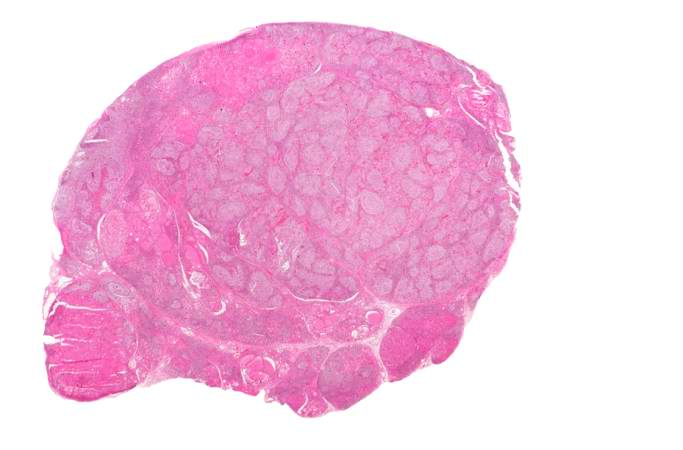
Revision as of 17:42, 20 August 2013 by Peter Anderson (talk | contribs) (This is a low-power photomicrograph of thyroid from this case. Note that the tissue is more cellular than one would expect and there does not appear to be normal colloid-filled blue spaces in this gland.)
No higher resolution available.
IPLab6Hashimoto2.jpg (683 × 450 pixels, file size: 27 KB, MIME type: image/jpeg)
This is a low-power photomicrograph of thyroid from this case. Note that the tissue is more cellular than one would expect and there does not appear to be normal colloid-filled blue spaces in this gland.
File history
Click on a date/time to view the file as it appeared at that time.
| Date/Time | Thumbnail | Dimensions | User | Comment | |
|---|---|---|---|---|---|
| current | 17:42, 20 August 2013 | | 683 × 450 (27 KB) | Peter Anderson (talk | contribs) | This is a low-power photomicrograph of thyroid from this case. Note that the tissue is more cellular than one would expect and there does not appear to be normal colloid-filled blue spaces in this gland. |
- You cannot overwrite this file.
File usage
There are no pages that link to this file.