File:IPLab4PulmonaryCongestion2.jpg
Revision as of 16:07, 19 August 2013 by Seung Park (talk | contribs) (This is a gross photograph of lung demonstrating acute pulmonary congestion and edema. A frothy exudate fills the bronchus (arrow).)
No higher resolution available.
IPLab4PulmonaryCongestion2.jpg (672 × 450 pixels, file size: 54 KB, MIME type: image/jpeg)
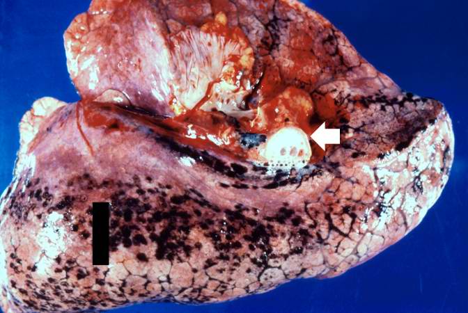
This is a gross photograph of lung demonstrating acute pulmonary congestion and edema. A frothy exudate fills the bronchus (arrow).
Pulmonary congestion is the engorgement of pulmonary vessels with blood. The increased pressure caused by this engorgement leads to transudation of fluid through the capillary walls and into the alveolar and interstitial spaces.
File history
Click on a date/time to view the file as it appeared at that time.
| Date/Time | Thumbnail | Dimensions | User | Comment | |
|---|---|---|---|---|---|
| current | 16:07, 19 August 2013 | | 672 × 450 (54 KB) | Seung Park (talk | contribs) | This is a gross photograph of lung demonstrating acute pulmonary congestion and edema. A frothy exudate fills the bronchus (arrow). |
- You cannot overwrite this file.
File usage
The following page links to this file: