File:IPLab3ChronicPepticUlcer10.jpg
Revision as of 04:17, 19 August 2013 by Seung Park (talk | contribs) (This is a photomicrograph of the serosal surface (1) from a section of stomach near the ulcer. Note that the inflammatory reaction extends out to the serosa.)
No higher resolution available.
IPLab3ChronicPepticUlcer10.jpg (679 × 450 pixels, file size: 65 KB, MIME type: image/jpeg)
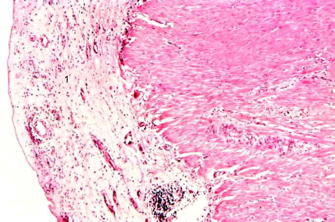
This is a photomicrograph of the serosal surface (1) from a section of stomach near the ulcer. Note that the inflammatory reaction extends out to the serosa.
File history
Click on a date/time to view the file as it appeared at that time.
| Date/Time | Thumbnail | Dimensions | User | Comment | |
|---|---|---|---|---|---|
| current | 04:17, 19 August 2013 | | 679 × 450 (65 KB) | Seung Park (talk | contribs) | This is a photomicrograph of the serosal surface (1) from a section of stomach near the ulcer. Note that the inflammatory reaction extends out to the serosa. |
- You cannot overwrite this file.
File usage
There are no pages that link to this file.