File:IPLab2FattyChange10.jpg
Revision as of 20:44, 19 June 2020 by Peter Anderson (talk | contribs) (Peter Anderson uploaded a new version of File:IPLab2FattyChange10.jpg)

Size of this preview: 800 × 436 pixels. Other resolutions: 320 × 174 pixels | 911 × 496 pixels.
Original file (911 × 496 pixels, file size: 672 KB, MIME type: image/jpeg)
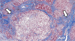
This is a low-power photomicrograph of liver stained with a trichrome stain. In this section, connective tissue stains green (arrows) and hepatic parenchymal cells are red. Note that many of the parenchymal cells have clear spaces indicating fatty degeneration. The proliferation of scar tissue between the liver lobules is the result of cirrhosis.
Cirrhosis is a liver disease characterized by necrosis, fibrosis, loss of normal liver architecture, and hyperplastic nodules.
File history
Click on a date/time to view the file as it appeared at that time.
| Date/Time | Thumbnail | Dimensions | User | Comment | |
|---|---|---|---|---|---|
| current | 20:44, 19 June 2020 | | 911 × 496 (672 KB) | Peter Anderson (talk | contribs) | |
| 17:00, 19 August 2013 | 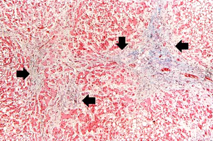 | 680 × 450 (86 KB) | Peter Anderson (talk | contribs) | This is a low-power photomicrograph of liver stained with a trichrome stain. In this section, connective tissue stains green (arrows) and hepatic parenchymal cells are red. Note that many of the parenchymal cells have clear spaces indicating fatty dege... |
- You cannot overwrite this file.
File usage
The following file is a duplicate of this file (more details):
There are no pages that link to this file.