File:IPLab2Atrophy4.jpg
Revision as of 16:03, 19 August 2013 by Peter Anderson (talk | contribs) (This is a higher-power photomicrograph indicating loss of testicular parenchymal tissue. There are very few recognizable spermatic cells in this tissue. The cluster of cells in the upper right is a focus of interstitial or Leydig cells (arrow). These c...)
No higher resolution available.
IPLab2Atrophy4.jpg (667 × 450 pixels, file size: 68 KB, MIME type: image/jpeg)
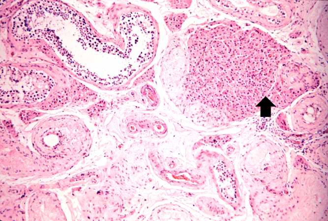
This is a higher-power photomicrograph indicating loss of testicular parenchymal tissue. There are very few recognizable spermatic cells in this tissue. The cluster of cells in the upper right is a focus of interstitial or Leydig cells (arrow). These cells are not affected by the hormone-induced atrophy process.
File history
Click on a date/time to view the file as it appeared at that time.
| Date/Time | Thumbnail | Dimensions | User | Comment | |
|---|---|---|---|---|---|
| current | 16:03, 19 August 2013 | | 667 × 450 (68 KB) | Peter Anderson (talk | contribs) | This is a higher-power photomicrograph indicating loss of testicular parenchymal tissue. There are very few recognizable spermatic cells in this tissue. The cluster of cells in the upper right is a focus of interstitial or Leydig cells (arrow). These c... |
- You cannot overwrite this file.
File usage
The following page links to this file: