File:IPLab13Myelomeningocele2.jpg
Revision as of 05:44, 21 August 2013 by Seung Park (talk | contribs) (This gross photograph shows consecutive lumbar vertebra from this case. Note the defect (arrows) in the two vertebral bodies on the right. This defect was caused by failure of the vertebral column to properly close.)
No higher resolution available.
IPLab13Myelomeningocele2.jpg (664 × 450 pixels, file size: 15 KB, MIME type: image/jpeg)
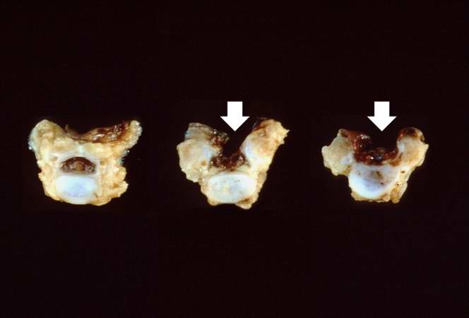
This gross photograph shows consecutive lumbar vertebra from this case. Note the defect (arrows) in the two vertebral bodies on the right. This defect was caused by failure of the vertebral column to properly close.
File history
Click on a date/time to view the file as it appeared at that time.
| Date/Time | Thumbnail | Dimensions | User | Comment | |
|---|---|---|---|---|---|
| current | 05:44, 21 August 2013 | | 664 × 450 (15 KB) | Seung Park (talk | contribs) | This gross photograph shows consecutive lumbar vertebra from this case. Note the defect (arrows) in the two vertebral bodies on the right. This defect was caused by failure of the vertebral column to properly close. |
- You cannot overwrite this file.
File usage
The following page links to this file: