File:IPLab9ARF1.jpg
No higher resolution available.
IPLab9ARF1.jpg (676 × 450 pixels, file size: 48 KB, MIME type: image/jpeg)
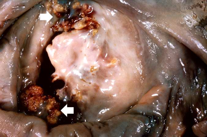
This is a gross photograph of mitral valve demonstrating marked thickening and fibrosis of the valve leaflet. There are also numerous foci of fibrinoid necrosis within the cusps and friable vegetations (verrucae) along the lines of closure (arrows). These irregular, warty projections are found at sites of erosion on the inflamed endocardial surface. The verrucae probably result from the precipitation of fibrin where the leaflets impinge on each other.
File history
Click on a date/time to view the file as it appeared at that time.
| Date/Time | Thumbnail | Dimensions | User | Comment | |
|---|---|---|---|---|---|
| current | 22:52, 20 August 2013 | | 676 × 450 (48 KB) | Seung Park (talk | contribs) | This is a gross photograph of mitral valve demonstrating marked thickening and fibrosis of the valve leaflet. There are also numerous foci of fibrinoid necrosis within the cusps and friable vegetations (verrucae) along the lines of closure (arrows). Th... |
You cannot overwrite this file.
File usage
The following page uses this file:
