File:IPLab9Diphtheria4.jpg
Revision as of 03:50, 21 August 2013 by Seung Park (talk | contribs) (In this higher-power photomicrograph of the tissue from the previous image, the ulcerated tracheal mucosa and the diphtheritic membrane are more clearly seen. Although difficult to make out at this magnification, most of the cells in this inflammatory ...)
No higher resolution available.
IPLab9Diphtheria4.jpg (675 × 450 pixels, file size: 69 KB, MIME type: image/jpeg)
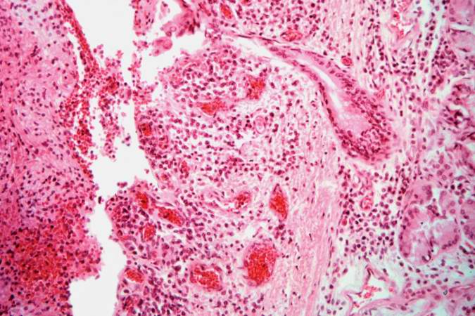
In this higher-power photomicrograph of the tissue from the previous image, the ulcerated tracheal mucosa and the diphtheritic membrane are more clearly seen. Although difficult to make out at this magnification, most of the cells in this inflammatory exudate are neutrophils.
File history
Click on a date/time to view the file as it appeared at that time.
| Date/Time | Thumbnail | Dimensions | User | Comment | |
|---|---|---|---|---|---|
| current | 03:50, 21 August 2013 | | 675 × 450 (69 KB) | Seung Park (talk | contribs) | In this higher-power photomicrograph of the tissue from the previous image, the ulcerated tracheal mucosa and the diphtheritic membrane are more clearly seen. Although difficult to make out at this magnification, most of the cells in this inflammatory ... |
- You cannot overwrite this file.
File usage
The following page links to this file: