File:IPLab7Melanoma7.jpg
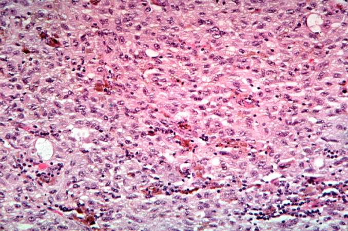
Revision as of 01:56, 21 August 2013 by Seung Park (talk | contribs) (This is a high-power photomicrograph of the main tumor mass with the cells growing as poorly formed nests and sheets of cells. There is little if any pigment in this section.)
No higher resolution available.
IPLab7Melanoma7.jpg (678 × 450 pixels, file size: 81 KB, MIME type: image/jpeg)
This is a high-power photomicrograph of the main tumor mass with the cells growing as poorly formed nests and sheets of cells. There is little if any pigment in this section.
File history
Click on a date/time to view the file as it appeared at that time.
| Date/Time | Thumbnail | Dimensions | User | Comment | |
|---|---|---|---|---|---|
| current | 01:56, 21 August 2013 | | 678 × 450 (81 KB) | Seung Park (talk | contribs) | This is a high-power photomicrograph of the main tumor mass with the cells growing as poorly formed nests and sheets of cells. There is little if any pigment in this section. |
- You cannot overwrite this file.
File usage
There are no pages that link to this file.