File:IPLab6ChronicRejection5.jpg
Revision as of 21:49, 20 August 2013 by Seung Park (talk | contribs) (This high-power photomicrograph of glomeruli from this kidney demonstrates congestion (1), increased cellularity of glomeruli with mesangial expansion, and a glomerulus that is almost completely obliterated or sclerosed (2).)
No higher resolution available.
IPLab6ChronicRejection5.jpg (672 × 450 pixels, file size: 71 KB, MIME type: image/jpeg)
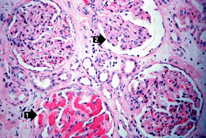
This high-power photomicrograph of glomeruli from this kidney demonstrates congestion (1), increased cellularity of glomeruli with mesangial expansion, and a glomerulus that is almost completely obliterated or sclerosed (2).
File history
Click on a date/time to view the file as it appeared at that time.
| Date/Time | Thumbnail | Dimensions | User | Comment | |
|---|---|---|---|---|---|
| current | 21:49, 20 August 2013 | | 672 × 450 (71 KB) | Seung Park (talk | contribs) | This high-power photomicrograph of glomeruli from this kidney demonstrates congestion (1), increased cellularity of glomeruli with mesangial expansion, and a glomerulus that is almost completely obliterated or sclerosed (2). |
- You cannot overwrite this file.
File usage
There are no pages that link to this file.