File:IPLab6ChronicRejection3.jpg
Revision as of 21:48, 20 August 2013 by Seung Park (talk | contribs) (This is a photomicrograph of kidney with a focal area of hemorrhage around a small blood vessel (left) and congestion of the glomeruli. Note that there is a marked loss of renal tubules throughout this section with replacement by fibrous connective tis...)
No higher resolution available.
IPLab6ChronicRejection3.jpg (681 × 450 pixels, file size: 89 KB, MIME type: image/jpeg)
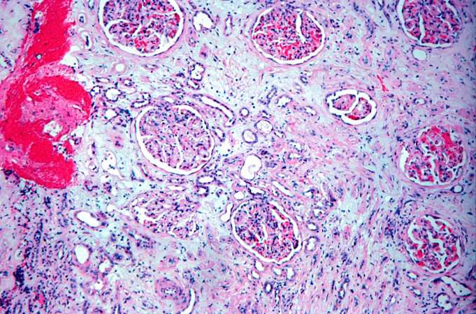
This is a photomicrograph of kidney with a focal area of hemorrhage around a small blood vessel (left) and congestion of the glomeruli. Note that there is a marked loss of renal tubules throughout this section with replacement by fibrous connective tissue. Also note the cellularity of the glomeruli.
File history
Click on a date/time to view the file as it appeared at that time.
| Date/Time | Thumbnail | Dimensions | User | Comment | |
|---|---|---|---|---|---|
| current | 21:48, 20 August 2013 | | 681 × 450 (89 KB) | Seung Park (talk | contribs) | This is a photomicrograph of kidney with a focal area of hemorrhage around a small blood vessel (left) and congestion of the glomeruli. Note that there is a marked loss of renal tubules throughout this section with replacement by fibrous connective tis... |
- You cannot overwrite this file.
File usage
There are no pages that link to this file.