File:IPLab6GN9.jpg
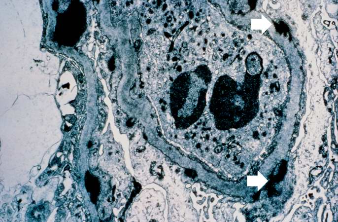
Revision as of 20:30, 20 August 2013 by Peter Anderson (talk | contribs) (This electron micrograph demonstrates scattered subepithelial dense deposits (arrows) and a polymorphonuclear leukocyte in the lumen.)
No higher resolution available.
IPLab6GN9.jpg (686 × 450 pixels, file size: 74 KB, MIME type: image/jpeg)
This electron micrograph demonstrates scattered subepithelial dense deposits (arrows) and a polymorphonuclear leukocyte in the lumen.
File history
Click on a date/time to view the file as it appeared at that time.
| Date/Time | Thumbnail | Dimensions | User | Comment | |
|---|---|---|---|---|---|
| current | 20:30, 20 August 2013 | | 686 × 450 (74 KB) | Peter Anderson (talk | contribs) | This electron micrograph demonstrates scattered subepithelial dense deposits (arrows) and a polymorphonuclear leukocyte in the lumen. |
- You cannot overwrite this file.
File usage
The following page links to this file: