File:IPLab6TB5.jpg
Revision as of 20:13, 20 August 2013 by Peter Anderson (talk | contribs) (High-power photomicrograph of a TB granuloma with multinucleated giant cells adjacent to an area of caseous necrosis (to the left).)
No higher resolution available.
IPLab6TB5.jpg (700 × 467 pixels, file size: 194 KB, MIME type: image/jpeg)
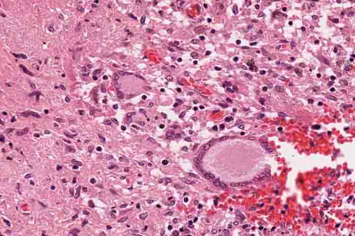
High-power photomicrograph of a TB granuloma with multinucleated giant cells adjacent to an area of caseous necrosis (to the left).
Caseous means cheesy.
File history
Click on a date/time to view the file as it appeared at that time.
| Date/Time | Thumbnail | Dimensions | User | Comment | |
|---|---|---|---|---|---|
| current | 20:13, 20 August 2013 | | 700 × 467 (194 KB) | Peter Anderson (talk | contribs) | High-power photomicrograph of a TB granuloma with multinucleated giant cells adjacent to an area of caseous necrosis (to the left). |
- You cannot overwrite this file.
File usage
The following page links to this file: