File:IPLab5DM3.jpg
No higher resolution available.
IPLab5DM3.jpg (675 × 450 pixels, file size: 94 KB, MIME type: image/jpeg)
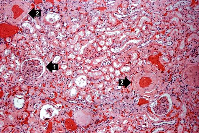
This is a higher-power photomicrograph of the cortical region. In this region there is ischemic obsolescence of glomeruli and one glomerulus with nodular glomerulosclerosis (1). Also note the thickened walls of the blood vessels (2).
File history
Click on a date/time to view the file as it appeared at that time.
| Date/Time | Thumbnail | Dimensions | User | Comment | |
|---|---|---|---|---|---|
| current | 10:27, 20 August 2013 | | 675 × 450 (94 KB) | Peter Anderson (talk | contribs) | This is a higher-power photomicrograph of the cortical region. In this region there is ischemic obsolescence of glomeruli and one glomerulus with nodular glomerulosclerosis (1). Also note the thickened walls of the blood vessels (2). |
You cannot overwrite this file.
File usage
There are no pages that use this file.
