File:IPLab5Hemochromatosis1.jpg
Revision as of 14:52, 20 August 2013 by Peter Anderson (talk | contribs) (This is a gross photograph of liver (1) and pancreas (2) from this case of hemochromatosis. Note that both of these organs have a dark brown coloration.)
No higher resolution available.
IPLab5Hemochromatosis1.jpg (688 × 450 pixels, file size: 59 KB, MIME type: image/jpeg)
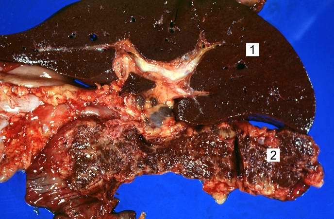
This is a gross photograph of liver (1) and pancreas (2) from this case of hemochromatosis. Note that both of these organs have a dark brown coloration.
File history
Click on a date/time to view the file as it appeared at that time.
| Date/Time | Thumbnail | Dimensions | User | Comment | |
|---|---|---|---|---|---|
| current | 14:52, 20 August 2013 | | 688 × 450 (59 KB) | Peter Anderson (talk | contribs) | This is a gross photograph of liver (1) and pancreas (2) from this case of hemochromatosis. Note that both of these organs have a dark brown coloration. |
- You cannot overwrite this file.
File usage
There are no pages that link to this file.