File:IPLab5Antitrypsin10.jpg
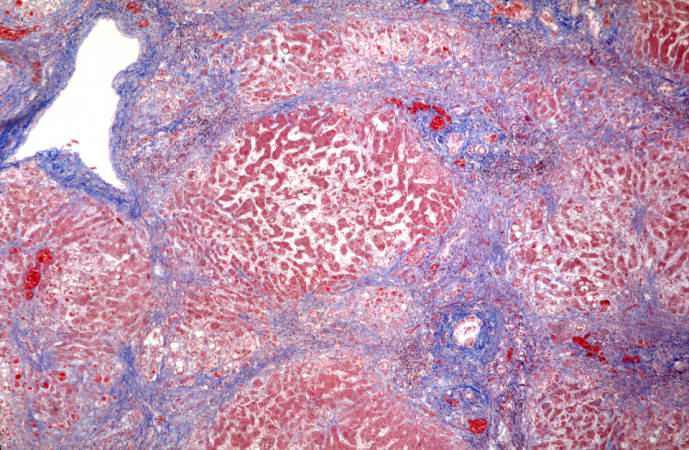
Revision as of 18:27, 19 August 2013 by Peter Anderson (talk | contribs) (This is a low-power photomicrograph of a trichrome-stained section of liver. There is bridging fibrosis (blue material) between portal regions.)
No higher resolution available.
IPLab5Antitrypsin10.jpg (689 × 450 pixels, file size: 80 KB, MIME type: image/jpeg)
This is a low-power photomicrograph of a trichrome-stained section of liver. There is bridging fibrosis (blue material) between portal regions.
File history
Click on a date/time to view the file as it appeared at that time.
| Date/Time | Thumbnail | Dimensions | User | Comment | |
|---|---|---|---|---|---|
| current | 18:27, 19 August 2013 | | 689 × 450 (80 KB) | Peter Anderson (talk | contribs) | This is a low-power photomicrograph of a trichrome-stained section of liver. There is bridging fibrosis (blue material) between portal regions. |
- You cannot overwrite this file.
File usage
The following page links to this file: