File:IPLab5Antitrypsin7.jpg
Revision as of 18:26, 19 August 2013 by Peter Anderson (talk | contribs) (This is a gross photograph of the cut section of liver from this case. In this view the liver looks smaller than normal and there is a definite micronodular appearance.)
No higher resolution available.
IPLab5Antitrypsin7.jpg (675 × 450 pixels, file size: 25 KB, MIME type: image/jpeg)
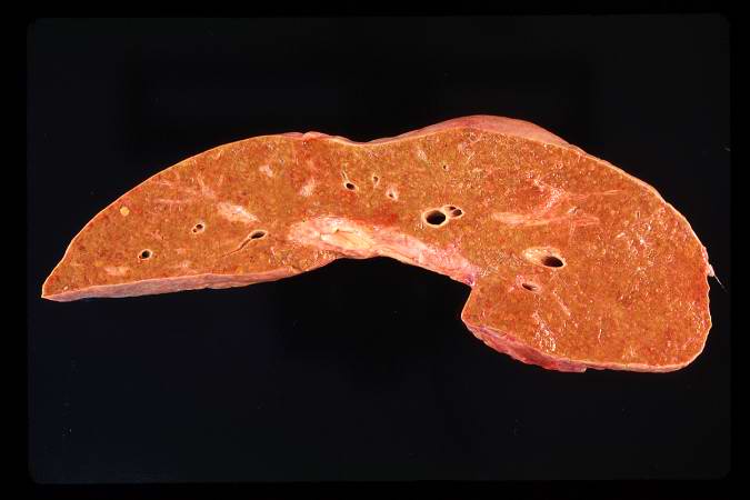
This is a gross photograph of the cut section of liver from this case. In this view the liver looks smaller than normal and there is a definite micronodular appearance.
File history
Click on a date/time to view the file as it appeared at that time.
| Date/Time | Thumbnail | Dimensions | User | Comment | |
|---|---|---|---|---|---|
| current | 18:26, 19 August 2013 | | 675 × 450 (25 KB) | Peter Anderson (talk | contribs) | This is a gross photograph of the cut section of liver from this case. In this view the liver looks smaller than normal and there is a definite micronodular appearance. |
- You cannot overwrite this file.
File usage
The following page links to this file: