File:IPLab5PolycysticKidney5.jpg
Revision as of 17:51, 19 August 2013 by Peter Anderson (talk | contribs) (This is another low-power photomicrograph of an H&E-stained section from this polycystic kidney. Again note the large cystic structures (arrows)and the fibrous connective tissue throughout this section.)
No higher resolution available.
IPLab5PolycysticKidney5.jpg (680 × 450 pixels, file size: 53 KB, MIME type: image/jpeg)
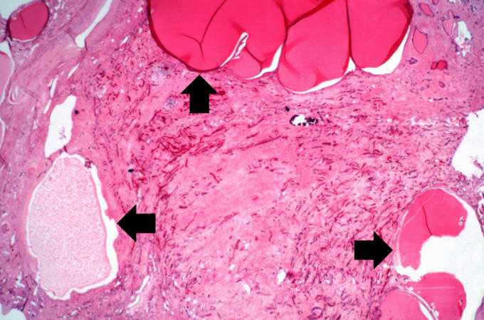
This is another low-power photomicrograph of an H&E-stained section from this polycystic kidney. Again note the large cystic structures (arrows)and the fibrous connective tissue throughout this section.
File history
Click on a date/time to view the file as it appeared at that time.
| Date/Time | Thumbnail | Dimensions | User | Comment | |
|---|---|---|---|---|---|
| current | 17:51, 19 August 2013 | | 680 × 450 (53 KB) | Peter Anderson (talk | contribs) | This is another low-power photomicrograph of an H&E-stained section from this polycystic kidney. Again note the large cystic structures (arrows)and the fibrous connective tissue throughout this section. |
- You cannot overwrite this file.
File usage
The following page links to this file: