File:IPLab5PolycysticKidney4.jpg
Revision as of 17:51, 19 August 2013 by Peter Anderson (talk | contribs) (This is a low-power photomicrograph of an H&E-stained section from this polycystic kidney. Note the large cystic structures (1), the few residual glomeruli (2), and the fibrous connective tissue throughout this section.)
No higher resolution available.
IPLab5PolycysticKidney4.jpg (684 × 450 pixels, file size: 69 KB, MIME type: image/jpeg)
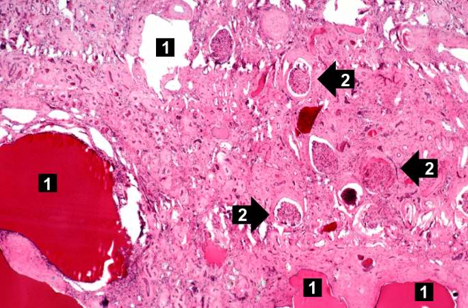
This is a low-power photomicrograph of an H&E-stained section from this polycystic kidney. Note the large cystic structures (1), the few residual glomeruli (2), and the fibrous connective tissue throughout this section.
File history
Click on a date/time to view the file as it appeared at that time.
| Date/Time | Thumbnail | Dimensions | User | Comment | |
|---|---|---|---|---|---|
| current | 17:51, 19 August 2013 | | 684 × 450 (69 KB) | Peter Anderson (talk | contribs) | This is a low-power photomicrograph of an H&E-stained section from this polycystic kidney. Note the large cystic structures (1), the few residual glomeruli (2), and the fibrous connective tissue throughout this section. |
- You cannot overwrite this file.
File usage
The following page links to this file: