File:IPLab5Neurofibromatosis2.jpg
No higher resolution available.
IPLab5Neurofibromatosis2.jpg (668 × 450 pixels, file size: 40 KB, MIME type: image/jpeg)
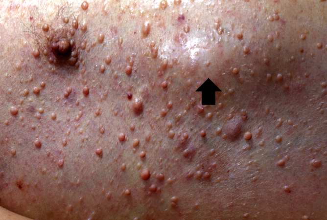
This is another view taken at autopsy demonstrating the neurofibromas. Some lesions can be seen as subcutaneous swellings (arrow) and others form pedunculated masses. Most are hyperpigmented.
File history
Click on a date/time to view the file as it appeared at that time.
| Date/Time | Thumbnail | Dimensions | User | Comment | |
|---|---|---|---|---|---|
| current | 12:19, 19 August 2013 | | 668 × 450 (40 KB) | Peter Anderson (talk | contribs) | This is another view taken at autopsy demonstrating the neurofibromas. Some lesions can be seen as subcutaneous swellings (arrow) and others form pedunculated masses. Most are hyperpigmented. |
You cannot overwrite this file.
File usage
There are no pages that use this file.
