File:IPLab2Atrophy8.jpg
Revision as of 16:11, 19 August 2013 by Peter Anderson (talk | contribs) (These kidneys were removed from a patient who had blockage of one ureter leading to increased pressure in the renal pelvis. The increased pressure produced hydronephrosis (arrow) in one kidney. What is the cause of atrophy in this case?)
No higher resolution available.
IPLab2Atrophy8.jpg (676 × 450 pixels, file size: 64 KB, MIME type: image/jpeg)
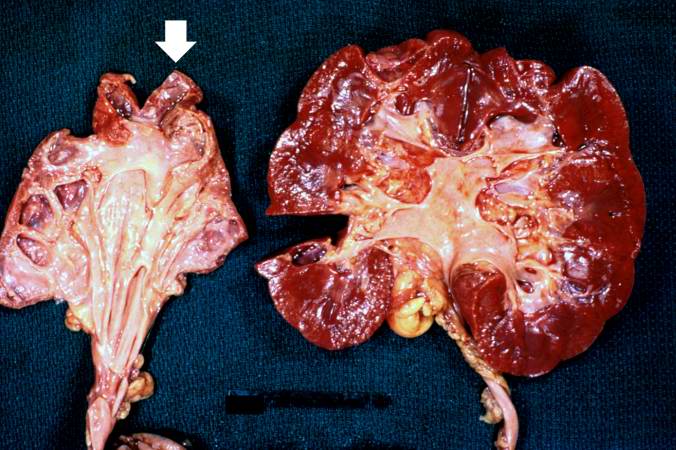
These kidneys were removed from a patient who had blockage of one ureter leading to increased pressure in the renal pelvis. The increased pressure produced hydronephrosis (arrow) in one kidney. What is the cause of atrophy in this case?
Hydronephrosis is dilation of the renal pelvis and atrophy of the cortex due to increase pressure from retained urine.
File history
Click on a date/time to view the file as it appeared at that time.
| Date/Time | Thumbnail | Dimensions | User | Comment | |
|---|---|---|---|---|---|
| current | 16:11, 19 August 2013 | | 676 × 450 (64 KB) | Peter Anderson (talk | contribs) | These kidneys were removed from a patient who had blockage of one ureter leading to increased pressure in the renal pelvis. The increased pressure produced hydronephrosis (arrow) in one kidney. What is the cause of atrophy in this case? |
- You cannot overwrite this file.
File usage
The following page links to this file: