File:IPLab2Hyperplasia5.jpg
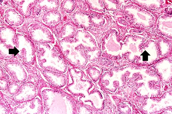
Revision as of 15:28, 19 August 2013 by Peter Anderson (talk | contribs) (Note these glands, which exhibit hyperplasia of the glandular epithelium. The infolding of the glandular epithelial cells forms papillary projections (arrows) into the lumen of the gland.)
No higher resolution available.
IPLab2Hyperplasia5.jpg (677 × 450 pixels, file size: 86 KB, MIME type: image/jpeg)
Note these glands, which exhibit hyperplasia of the glandular epithelium. The infolding of the glandular epithelial cells forms papillary projections (arrows) into the lumen of the gland.
File history
Click on a date/time to view the file as it appeared at that time.
| Date/Time | Thumbnail | Dimensions | User | Comment | |
|---|---|---|---|---|---|
| current | 15:28, 19 August 2013 | | 677 × 450 (86 KB) | Peter Anderson (talk | contribs) | Note these glands, which exhibit hyperplasia of the glandular epithelium. The infolding of the glandular epithelial cells forms papillary projections (arrows) into the lumen of the gland. |
- You cannot overwrite this file.
File usage
There are no pages that link to this file.