File:IPLab1KidneyInfarction6.jpg


Size of this preview: 800 × 459 pixels. Other resolution: 897 × 515 pixels.
Original file (897 × 515 pixels, file size: 258 KB, MIME type: image/jpeg)
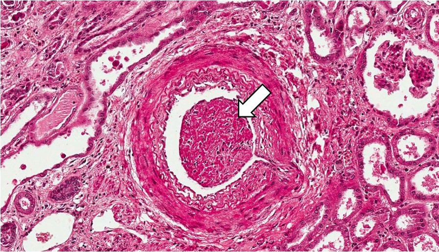
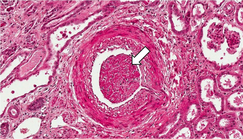
This section, taken at the cortical medullary junction, illustrates a blood vessel in the upper right portion of the slide (arrow) which is filled with thrombotic material. This vessel has occluded an end artery resulting in ischemia and infarction.
File history
Click on a date/time to view the file as it appeared at that time.
| Date/Time | Thumbnail | Dimensions | User | Comment | |
|---|---|---|---|---|---|
| current | 21:22, 27 June 2019 | | 897 × 515 (258 KB) | Peter Anderson (talk | contribs) | |
| 15:12, 15 August 2013 |  | 676 × 450 (64 KB) | Seung Park (talk | contribs) |
You cannot overwrite this file.
File usage
The following page uses this file:
