File:IPLab1Tuberculosis2.jpg
Revision as of 02:51, 16 August 2013 by Seung Park (talk | contribs)
No higher resolution available.
IPLab1Tuberculosis2.jpg (671 × 450 pixels, file size: 72 KB, MIME type: image/jpeg)
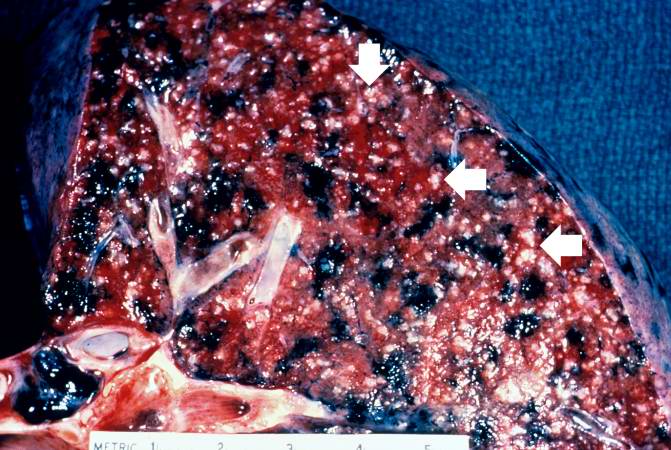
This is a closer view of the same section of lung containing multiple white granulomas which are now more easily identified (arrows). These lesions are referred to as miliary tuberculosis. Dark areas of anthracosis are also prominent in this lung.
Disseminated tuberculosis refers to the hematogenous spread of tuberculous lesions throughout the body. It is also known as miliary tuberculosis (which is so-called because the lesions resemble millet).
File history
Click on a date/time to view the file as it appeared at that time.
| Date/Time | Thumbnail | Dimensions | User | Comment | |
|---|---|---|---|---|---|
| current | 02:49, 16 August 2013 | | 671 × 450 (72 KB) | Seung Park (talk | contribs) |
- You cannot overwrite this file.
File usage
The following page links to this file: