File:IPLab13Hyaline9.jpg
No higher resolution available.
IPLab13Hyaline9.jpg (672 × 450 pixels, file size: 78 KB, MIME type: image/jpeg)
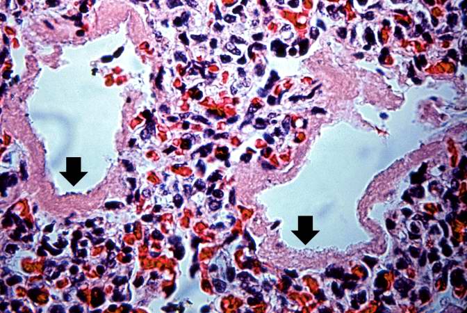
This higher-power photomicrograph shows more clearly the hyaline membranes (arrows) and the congestion in the interstitium.
File history
Click on a date/time to view the file as it appeared at that time.
| Date/Time | Thumbnail | Dimensions | User | Comment | |
|---|---|---|---|---|---|
| current | 00:52, 21 August 2013 | | 672 × 450 (78 KB) | Seung Park (talk | contribs) | This higher-power photomicrograph shows more clearly the hyaline membranes (arrows) and the congestion in the interstitium. |
You cannot overwrite this file.
File usage
The following page uses this file:
