File:IPLab12Alcoholic11.jpg
No higher resolution available.
IPLab12Alcoholic11.jpg (608 × 450 pixels, file size: 58 KB, MIME type: image/jpeg)
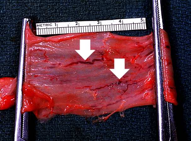
This photograph taken from still another patient at autopsy demonstrates the esophageal varices in the distal esophagus (arrows). The esophagus was clamped before removing the esophagus from the body in order to trap the blood in these distended varices. It is obvious how easily these thin-walled superficial varices could rupture and bleed.
File history
Click on a date/time to view the file as it appeared at that time.
| Date/Time | Thumbnail | Dimensions | User | Comment | |
|---|---|---|---|---|---|
| current | 00:16, 21 August 2013 | | 608 × 450 (58 KB) | Seung Park (talk | contribs) | This photograph taken from still another patient at autopsy demonstrates the esophageal varices in the distal esophagus (arrows). The esophagus was clamped before removing the esophagus from the body in order to trap the blood in these distended varice... |
You cannot overwrite this file.
File usage
The following page uses this file:
