File:IPLab10Mucor3.jpg
Revision as of 04:19, 21 August 2013 by Seung Park (talk | contribs) (This is an even higher-power photomicrograph of the wall of the carotid artery (1) and the thrombus (2). Within the wall of the artery and in the thrombus there are multiple variably shaped clear areas (3). At this magnification and with this stain, it...)
No higher resolution available.
IPLab10Mucor3.jpg (680 × 450 pixels, file size: 70 KB, MIME type: image/jpeg)
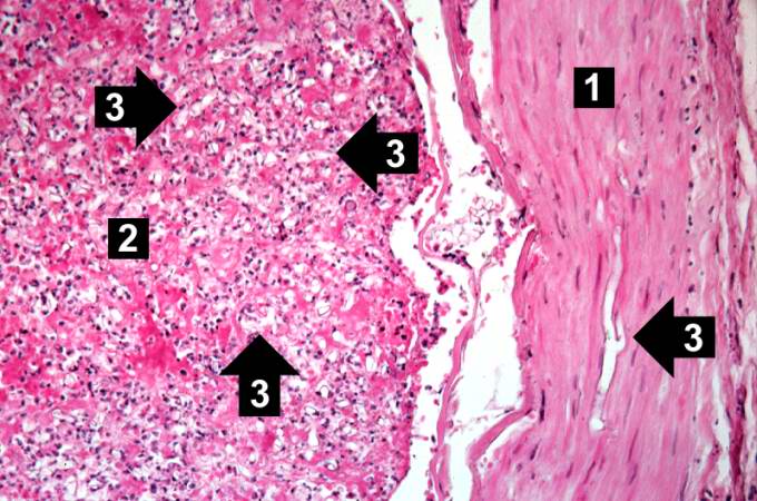
This is an even higher-power photomicrograph of the wall of the carotid artery (1) and the thrombus (2). Within the wall of the artery and in the thrombus there are multiple variably shaped clear areas (3). At this magnification and with this stain, it is impossible to determine what these clear spaces represent.
A thrombus is a solid mass resulting from the aggregation of blood constituents within the vascular system.
File history
Click on a date/time to view the file as it appeared at that time.
| Date/Time | Thumbnail | Dimensions | User | Comment | |
|---|---|---|---|---|---|
| current | 04:19, 21 August 2013 | | 680 × 450 (70 KB) | Seung Park (talk | contribs) | This is an even higher-power photomicrograph of the wall of the carotid artery (1) and the thrombus (2). Within the wall of the artery and in the thrombus there are multiple variably shaped clear areas (3). At this magnification and with this stain, it... |
- You cannot overwrite this file.
File usage
The following page links to this file: