File:IPLab10Blasto6.jpg
Revision as of 04:16, 21 August 2013 by Seung Park (talk | contribs) (This high-power photomicrograph shows what appear to be inflammatory cells filling the alveoli. At this magnification, numerous round bodies (arrows) that look like inflammatory cell nuclei can be seen. However, on closer examination, some of these rou...)
No higher resolution available.
IPLab10Blasto6.jpg (656 × 450 pixels, file size: 103 KB, MIME type: image/jpeg)
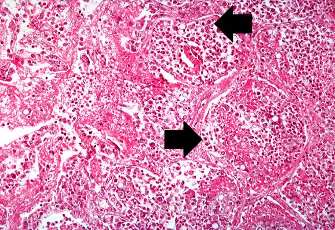
This high-power photomicrograph shows what appear to be inflammatory cells filling the alveoli. At this magnification, numerous round bodies (arrows) that look like inflammatory cell nuclei can be seen. However, on closer examination, some of these round bodies are surrounded by clear halos.
File history
Click on a date/time to view the file as it appeared at that time.
| Date/Time | Thumbnail | Dimensions | User | Comment | |
|---|---|---|---|---|---|
| current | 04:16, 21 August 2013 | | 656 × 450 (103 KB) | Seung Park (talk | contribs) | This high-power photomicrograph shows what appear to be inflammatory cells filling the alveoli. At this magnification, numerous round bodies (arrows) that look like inflammatory cell nuclei can be seen. However, on closer examination, some of these rou... |
- You cannot overwrite this file.
File usage
The following page links to this file: