Uploads by Peter Anderson
Jump to navigation
Jump to search
This special page shows all uploaded files.


| Date | Name | Thumbnail | Size | Description | Versions |
|---|---|---|---|---|---|
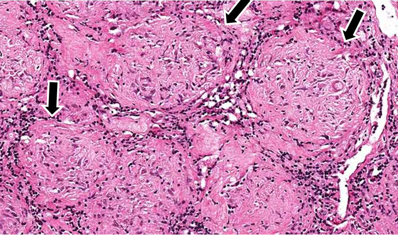
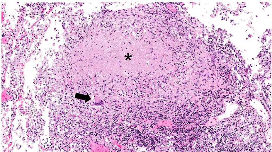
| 23:17, 19 June 2020 | IPLab3Sarcoidosis3b.jpg (file) |  |
520 KB | 1 | |
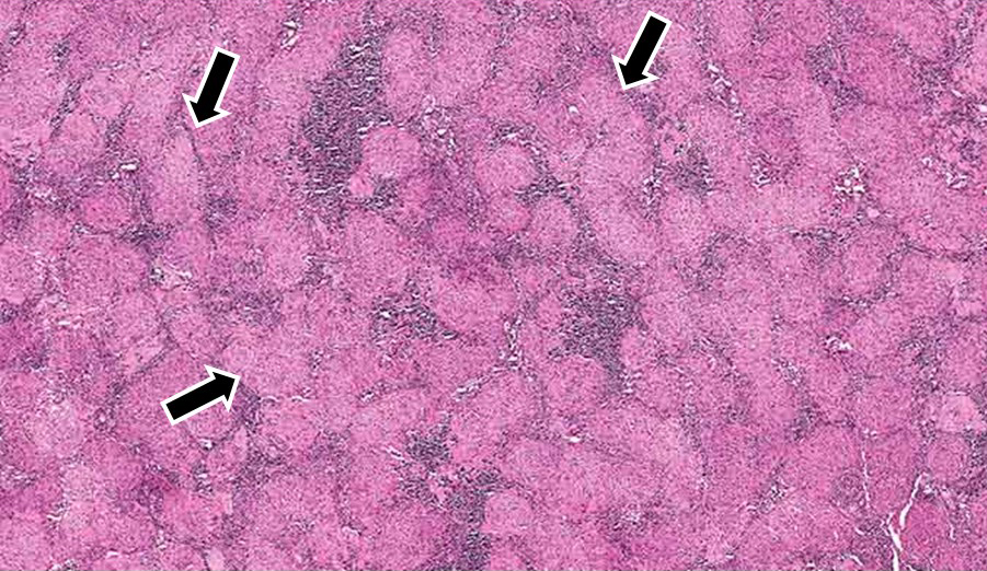
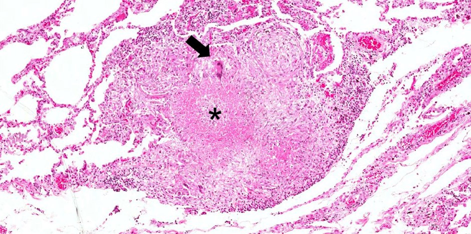
| 23:17, 19 June 2020 | IPLab3Sarcoidosis2b.jpg (file) |  |
452 KB | 1 | |
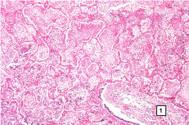
| 23:06, 19 June 2020 | IPLab3Bronchopneumonia6b.jpg (file) |  |
485 KB | 1 | |
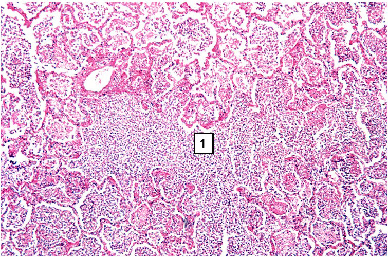
| 23:06, 19 June 2020 | IPLab3Bronchopneumonia5b.jpg (file) |  |
467 KB | 1 | |
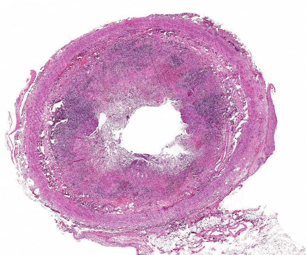
| 22:49, 19 June 2020 | IPLab3AcuteAppendicitis2x.jpg (file) |  |
496 KB | 1 | |
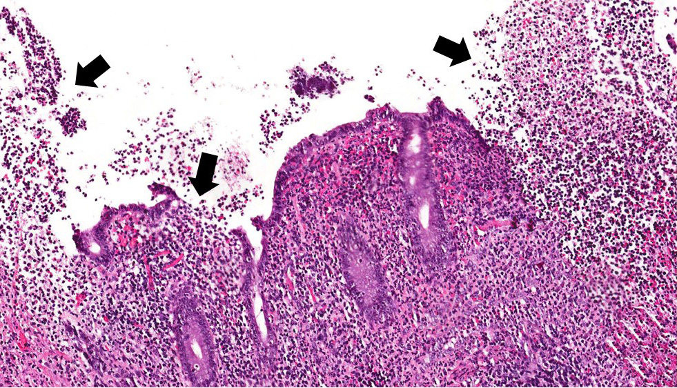
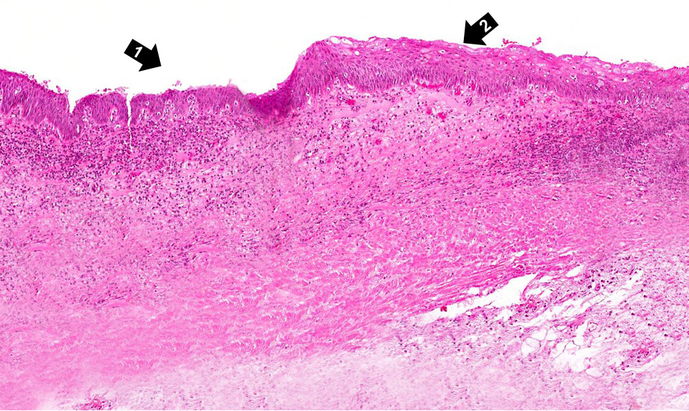
| 22:45, 19 June 2020 | IPLab3AcuteAppendicitis5b.jpg (file) |  |
463 KB | 1 | |
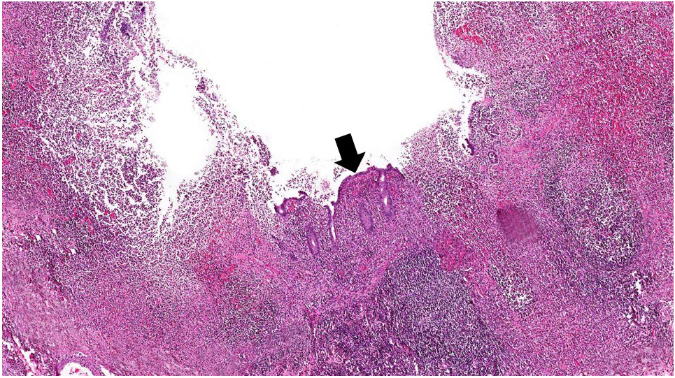
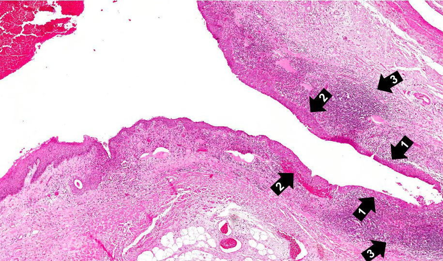
| 22:45, 19 June 2020 | IPLab3AcuteAppendicitis4b.jpg (file) |  |
441 KB | 1 | |
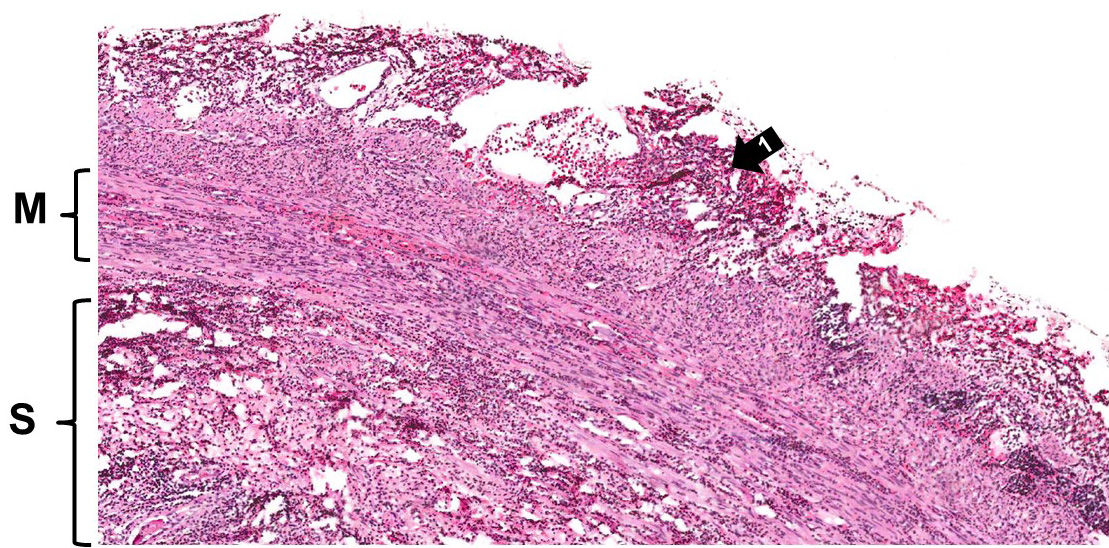
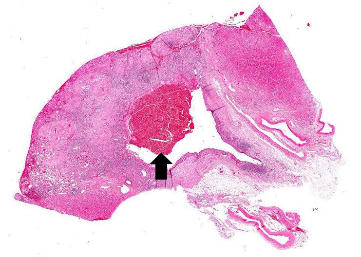
| 22:44, 19 June 2020 | IPLab3AcuteAppendicitis3b.jpg (file) |  |
418 KB | 1 | |
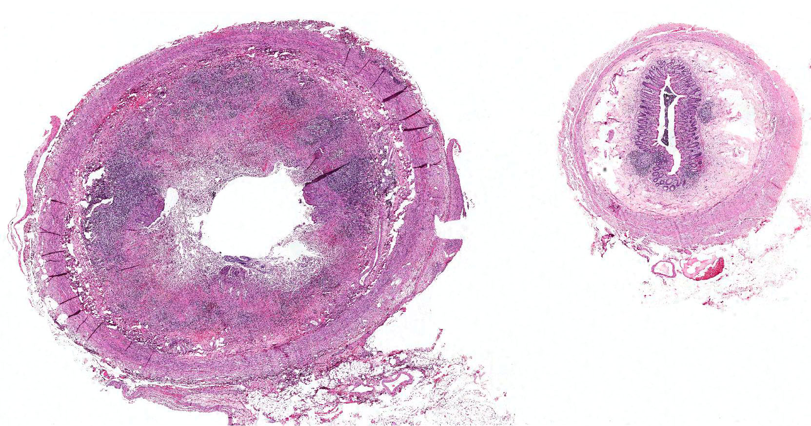
| 22:42, 19 June 2020 | IPLab3AcuteAppendicitis2b.jpg (file) |  |
520 KB | 1 | |
| 21:08, 19 June 2020 | IPLab2Metaplasia5b.jpg (file) |  |
571 KB | 1 | |
| 21:08, 19 June 2020 | IPLab2Metaplasia4b.jpg (file) |  |
727 KB | 1 | |
| 21:08, 19 June 2020 | IPLab2Metaplasia3b.jpg (file) |  |
633 KB | 1 | |
| 21:07, 19 June 2020 | IPLab2Metaplasia2b.JPG (file) |  |
329 KB | 1 | |
| 21:07, 19 June 2020 | IPLab2Metaplasia1b.JPG (file) |  |
442 KB | 1 | |
| 20:56, 19 June 2020 | IPLab2FattyChange5b.jpg (file) |  |
436 KB | 1 | |
| 20:55, 19 June 2020 | IPLab2FattyChange4b.jpg (file) |  |
667 KB | 1 | |
| 20:54, 19 June 2020 | IPLab2FattyChange3b.jpg (file) |  |
684 KB | 1 | |
| 20:51, 19 June 2020 | IPLab2FattyChange10b.jpg (file) |  |
672 KB | 1 | |
| 20:44, 19 June 2020 | IPLab2FattyChange10.jpg (file) |  |
672 KB | 2 | |
| 22:06, 27 June 2019 | IPLab1Tuberculosis7b.jpg (file) |  |
258 KB | 1 | |
| 22:06, 27 June 2019 | IPLab1Tuberculosis6b.jpg (file) |  |
200 KB | 1 | |
| 22:05, 27 June 2019 | IPLab1Tuberculosis5b.jpg (file) |  |
260 KB | 1 | |
| 22:04, 27 June 2019 | IPLab1Tuberculosis4b.jpg (file) |  |
172 KB | 1 | |
| 21:58, 27 June 2019 | IPLab1FatNecrosis10.jpg (file) |  |
166 KB | 1 | |
| 21:40, 27 June 2019 | IPLab1FatNecrosis8.jpg (file) |  |
166 KB | 2 | |
| 21:22, 27 June 2019 | IPLab1KidneyInfarction6.jpg (file) |  |
258 KB | 2 | |
| 21:20, 27 June 2019 | IPLab1KidneyInfarction5.jpg (file) |  |
242 KB | 2 | |
| 21:15, 27 June 2019 | IPLab1KidneyInfarction4.jpg (file) |  |
228 KB | 2 | |
| 18:55, 31 October 2016 | IPLab1Tuberculosis8b.jpg (file) |  |
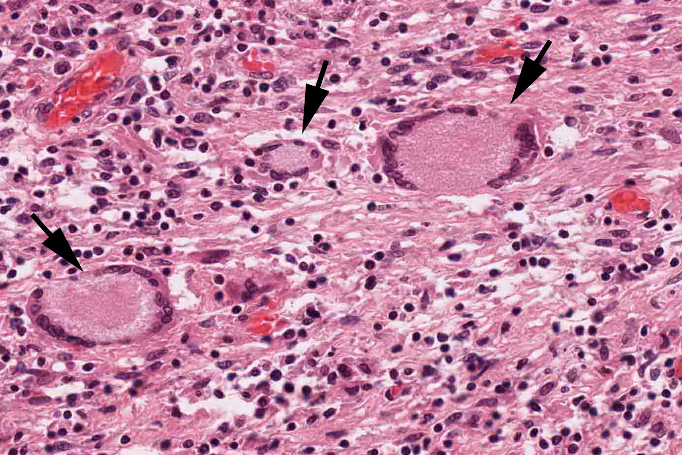
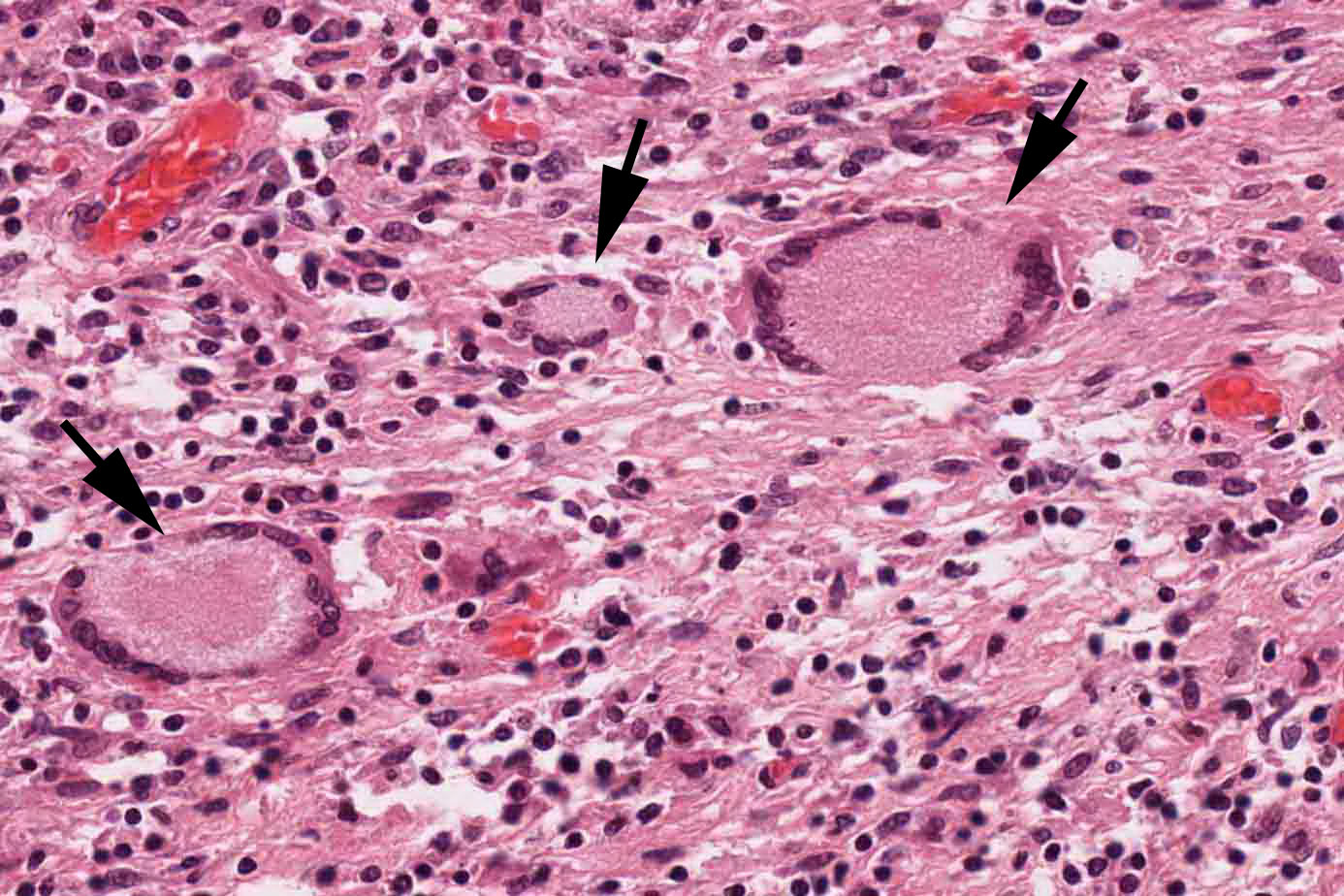
246 KB | This is a high-power photomicrograph of Langhans-type multinucleated giant cells (arrows) that are characteristic of tuberculous granulomas. Note the ring of the nuclei in these giant cells. | 1 |
| 18:42, 31 October 2016 | IPLab1Tuberculosis8.jpg (file) |  |
246 KB | 1 | |
| 22:27, 4 September 2013 | IPLab2Hypertrophy4.jpg (file) |  |
125 KB | 2 | |
| 22:26, 4 September 2013 | IPLab2Hypertrophy3.jpg (file) |  |
163 KB | 2 | |
| 22:24, 4 September 2013 | IPLab2Hypertrophy2.jpg (file) |  |
204 KB | 2 | |
| 20:43, 20 August 2013 | IPLab6GN1.jpg (file) |  |
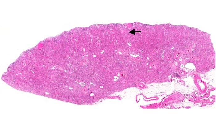
72 KB | This is a low-power photomicrograph of a saggital section of end stage chronic glomerulonephritis (GN). Note the marked thinning of the cortex (arrow). | 1 |
| 20:30, 20 August 2013 | IPLab6GN10.jpg (file) |  |
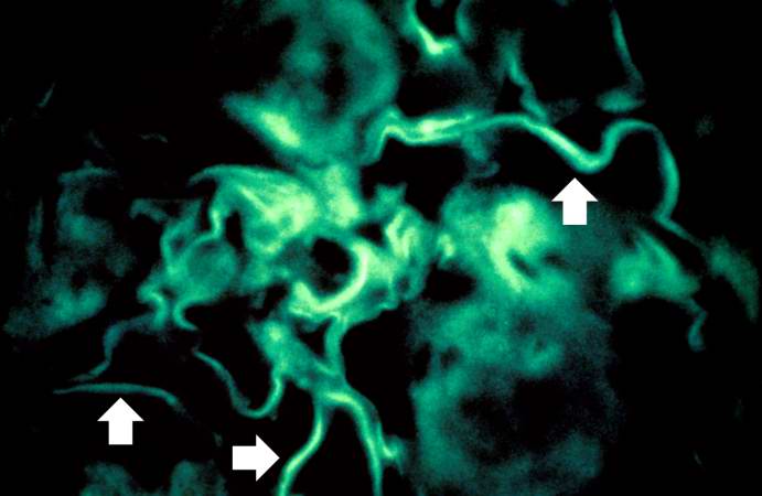
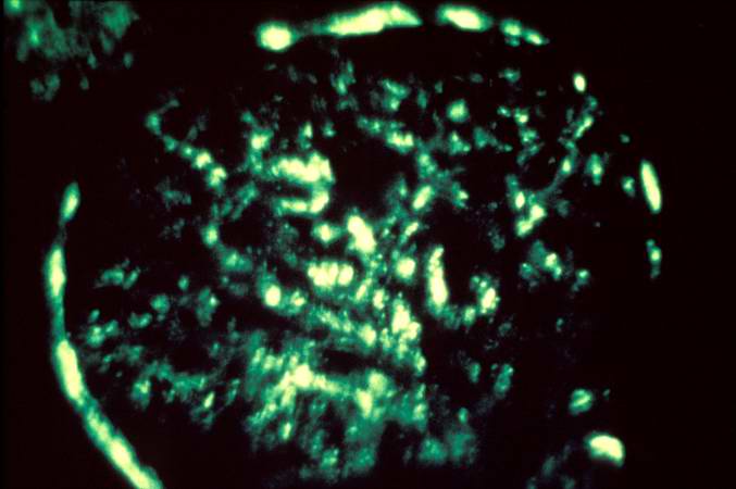
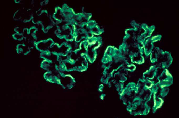
29 KB | For comparison this is an immunofluorescent photomicrograph of a glomerulus from a patient with Goodpasture's syndrome. The linear (arrows) immunofluorescence is characteristic of Goodpasture's syndrome. | 1 |
| 20:30, 20 August 2013 | IPLab6GN9.jpg (file) |  |
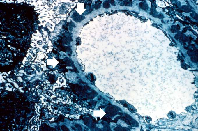
74 KB | This electron micrograph demonstrates scattered subepithelial dense deposits (arrows) and a polymorphonuclear leukocyte in the lumen. | 1 |
| 20:30, 20 August 2013 | IPLab6GN8.jpg (file) |  |
31 KB | This immunofluorescent photomicrograph of a glomerulus from a case of acute poststreptococcal glomerulonephritis shows a granular immunofluorescence pattern consistent with immune complex disease. The primary antibody used for this staining was specifi... | 1 |
| 20:29, 20 August 2013 | IPLab6GN7.jpg (file) |  |
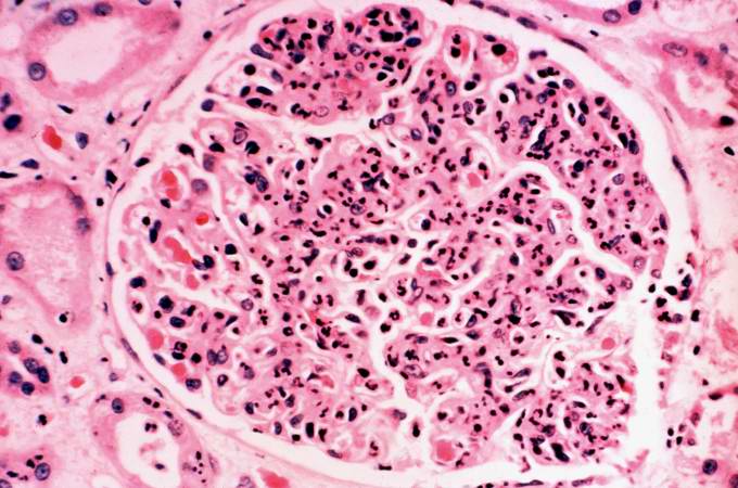
62 KB | This is a photomicrograph of a glomerulus from another case with acute poststreptococcal glomerulonephritis. In this case the immune complex glomerular disease is ongoing with necrosis and accumulation of neutrophils in the glomerulus. | 1 |
| 20:29, 20 August 2013 | IPLab6GN6.jpg (file) |  |
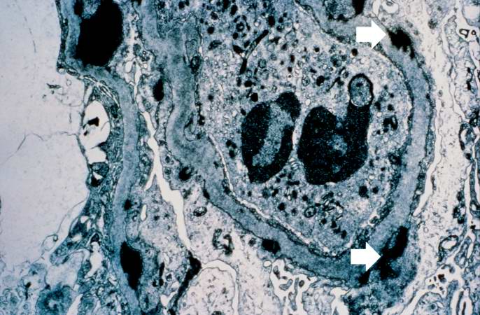
66 KB | This is an electron micrograph of subepithelial granular electron dense deposits (arrows) which correspond to the granular immunofluorescence seen in the previous image. | 1 |
| 20:28, 20 August 2013 | IPLab6GN5.jpg (file) |  |
30 KB | This is an immunofluorescent photomicrograph of granular membranous immunofluorescence (immune complex disease). The antibody used for these studies was specific for IgG. | 1 |
| 20:28, 20 August 2013 | IPLab6GN4.jpg (file) |  |
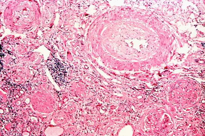
87 KB | This is a photomicrograph of interstitial and vascular lesions in end stage renal disease. | 1 |
| 20:27, 20 August 2013 | IPLab6GN3.jpg (file) |  |
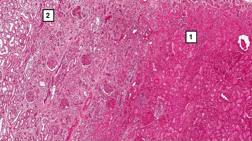
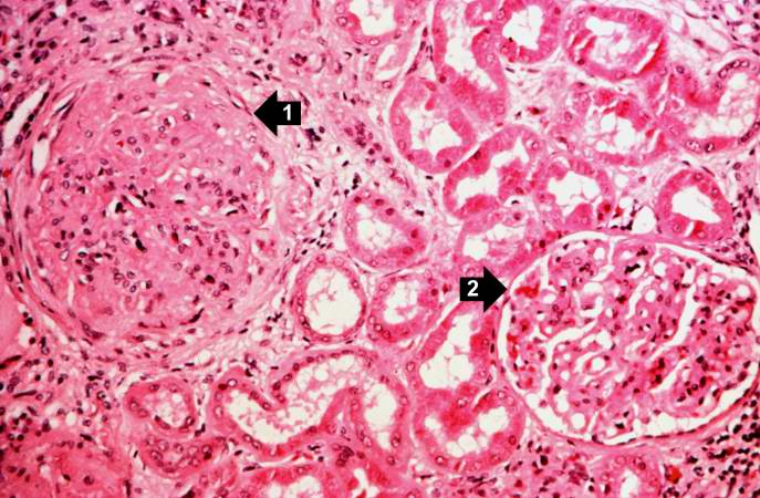
71 KB | This is a higher-power photomicrograph of hyalinized glomeruli (1) and glomeruli with thickened basement membranes (2). | 1 |
| 20:27, 20 August 2013 | IPLab6GN2.jpg (file) |  |
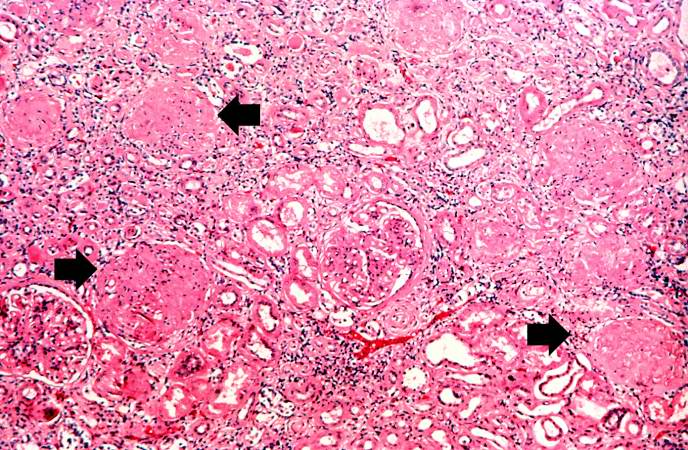
103 KB | This is a higher-power photomicrograph of hyalinized glomeruli (arrows) and glomeruli with thick basement membranes. | 1 |
| 20:14, 20 August 2013 | IPLab6TB6.jpg (file) |  |
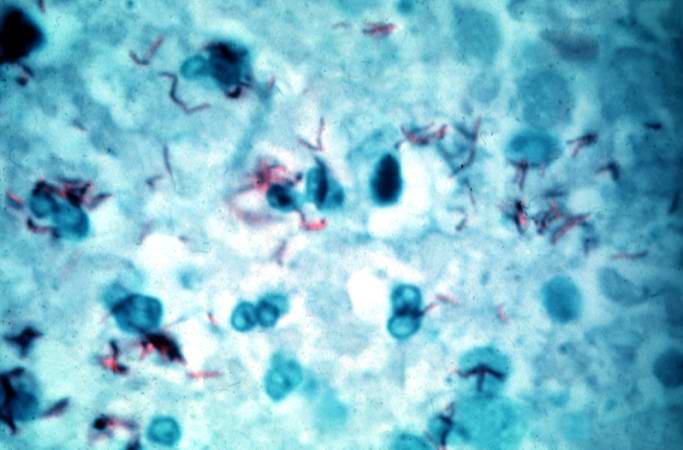
37 KB | This is a high-power (oil immersion) photomicrograph of granuloma stained with an acid-fast stain. Mycobacterium tuberculosis bacilli stain red. | 1 |
| 20:13, 20 August 2013 | IPLab6TB5.jpg (file) |  |
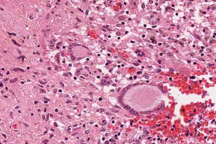
194 KB | High-power photomicrograph of a TB granuloma with multinucleated giant cells adjacent to an area of caseous necrosis (to the left). | 1 |
| 20:11, 20 August 2013 | IPLab6TB4.jpg (file) |  |
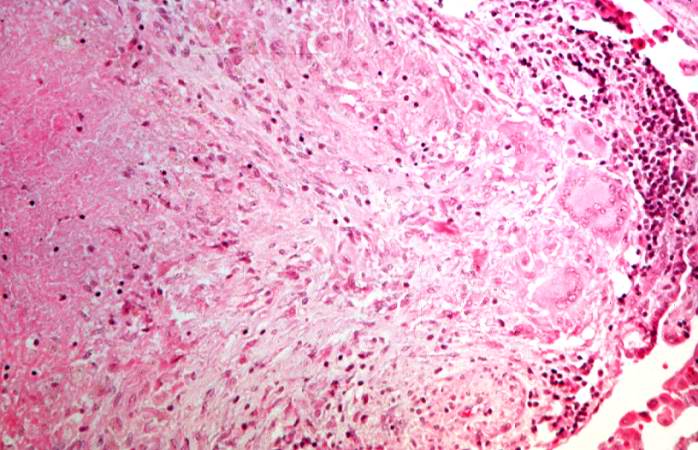
68 KB | This is a higher-power photomicrograph of a TB granuloma. The area of caseous necrosis is on the left side of the image, there are multinucleated giant cells and epithelioid macrophages throughout the remainder of the tissue. | 1 |
| 20:10, 20 August 2013 | IPLab6TB3.jpg (file) |  |
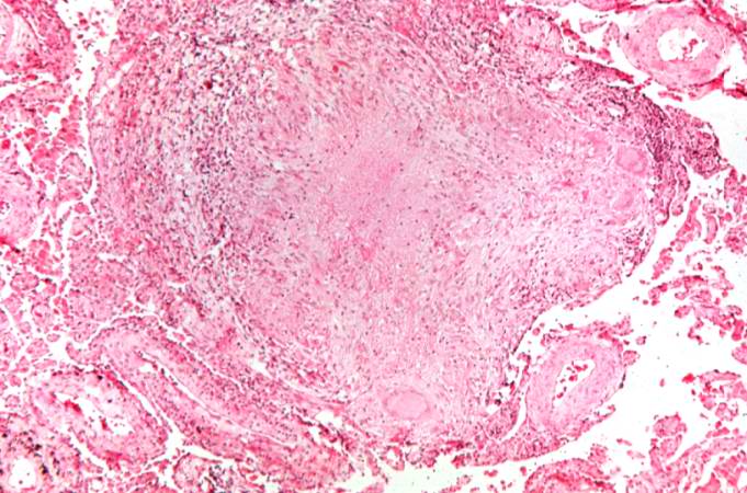
72 KB | This is a higher-power photomicrograph of a TB granuloma. Note the eosinophilic material in the center of this granuloma (caseous necrosis) and the epithelioid macrophages and giant cells around the periphery. | 1 |
| 20:10, 20 August 2013 | IPLab6TB2.jpg (file) |  |
36 KB | This is a low-power photomicrograph of lung tissue with multiple circumscribed nodules - granulomas (arrows). | 1 |
| 20:10, 20 August 2013 | IPLab6TB1.jpg (file) |  |
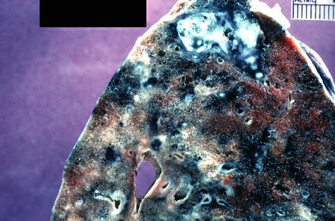
63 KB | This is a photograph of a section of lung with an apical lesion. This lesion represents an old healed lesion from Mycobacterium tuberculosis infection. | 1 |
| 19:59, 20 August 2013 | IPLab6Scleroderma5.jpg (file) |  |
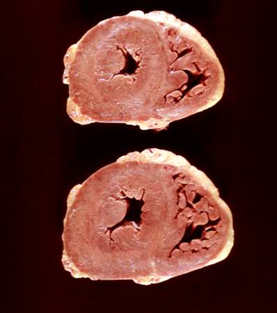
19 KB | This is a gross photograph of the heart from this case. There is thickening of the left ventricular wall and some thickening of the right ventricle as well. | 1 |