File list
This special page shows all uploaded files.
| Date | Name | Thumbnail | Size | User | Description | Versions |
|---|---|---|---|---|---|---|
| 02:37, 21 August 2013 | IPLab8HSVEncephalitis12.jpg (file) |  |
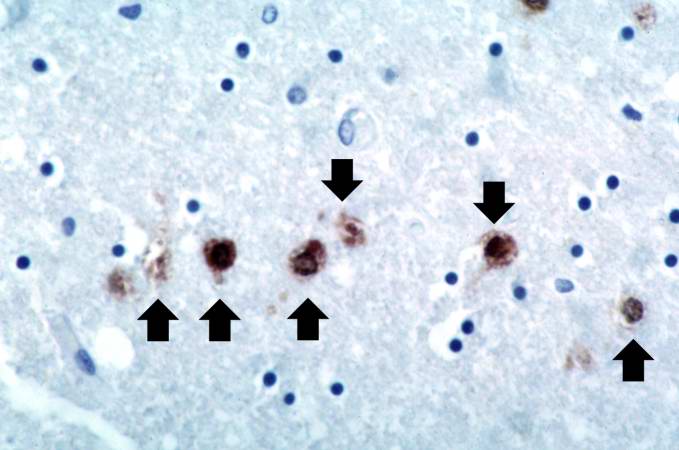
36 KB | Seung Park | This is a photomicrograph of a brain section stained with an antibody against herpes simplex. Even at this magnification, it is easy to pick out cells that are positive for the virus (arrows). | 1 |
| 02:37, 21 August 2013 | IPLab8HSVEncephalitis11.jpg (file) |  |
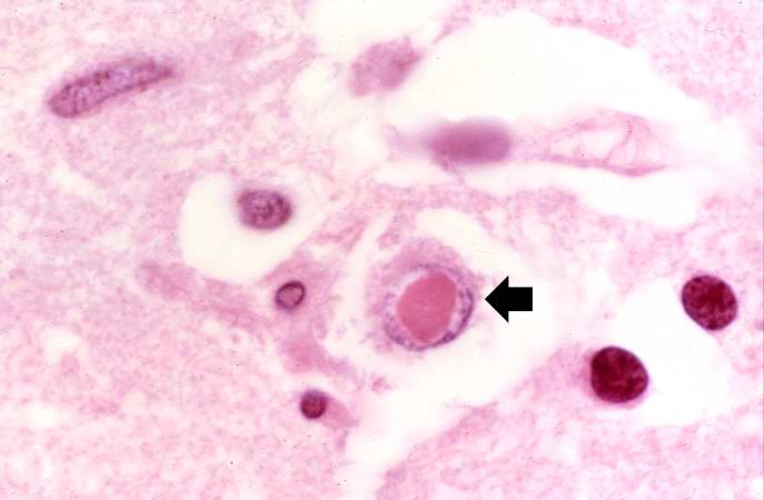
27 KB | Seung Park | This is another high-power photomicrograph of a cell containing an intranuclear inclusion body (arrow). | 1 |
| 02:37, 21 August 2013 | IPLab8HSVEncephalitis10.jpg (file) |  |
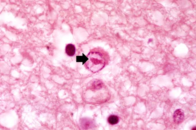
40 KB | Seung Park | This is another high-power photomicrograph of a cell containing an intranuclear inclusion body (arrow). Note that the nuclear chromatin has been pushed to the outer edges of the nucleus. | 1 |
| 02:36, 21 August 2013 | IPLab8HSVEncephalitis9.jpg (file) |  |
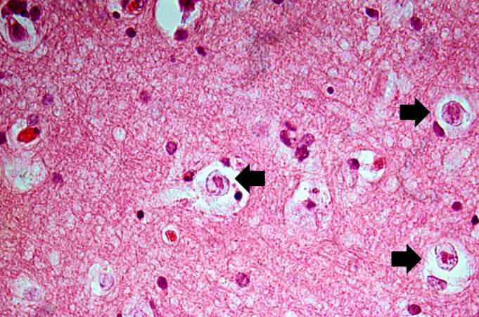
78 KB | Seung Park | This is a high-power photomicrograph demonstrating cells containing intranuclear inclusion bodies (arrows). | 1 |
| 02:36, 21 August 2013 | IPLab8HSVEncephalitis8.jpg (file) |  |
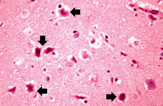
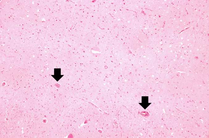
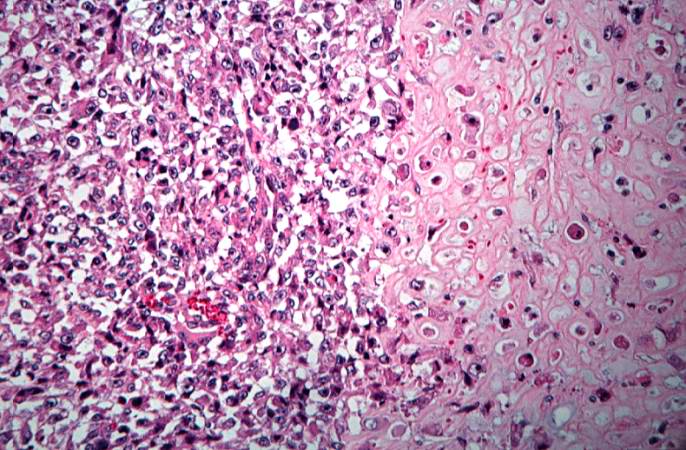
55 KB | Seung Park | This is a high-power photomicrograph showing several necrotic cells (arrows). | 1 |
| 02:36, 21 August 2013 | IPLab8HSVEncephalitis7.jpg (file) |  |
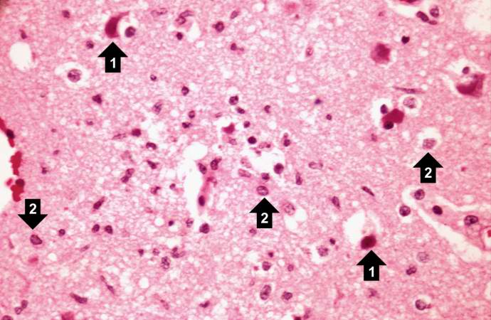
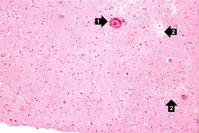
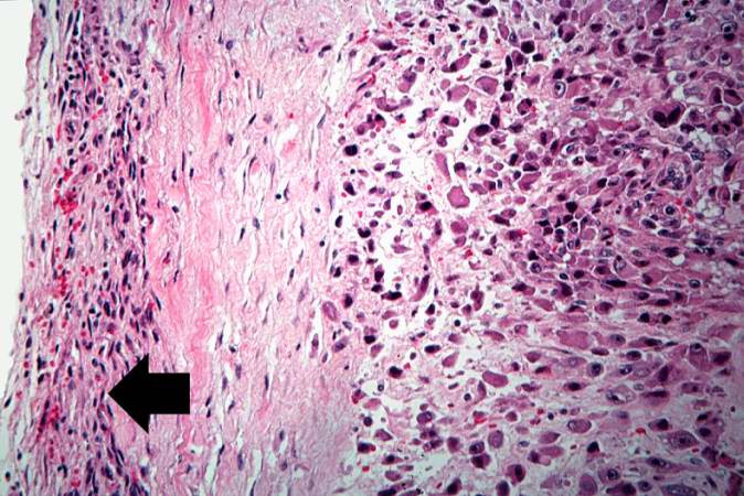
55 KB | Seung Park | This is a high-power photomicrograph demonstrating clear areas, which indicate edema, and numerous shrunken red necrotic cells (1). At this power, it can be seen that eosinophilic intranuclear inclusion bodies have displaced chromatin to the periphery ... | 1 |
| 02:35, 21 August 2013 | IPLab8HSVEncephalitis6.jpg (file) |  |
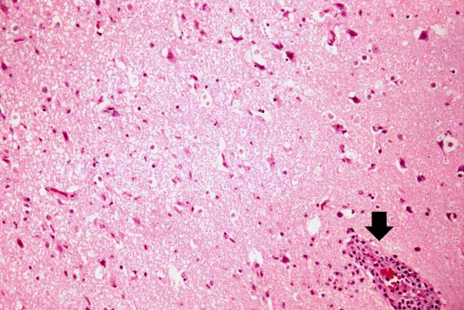
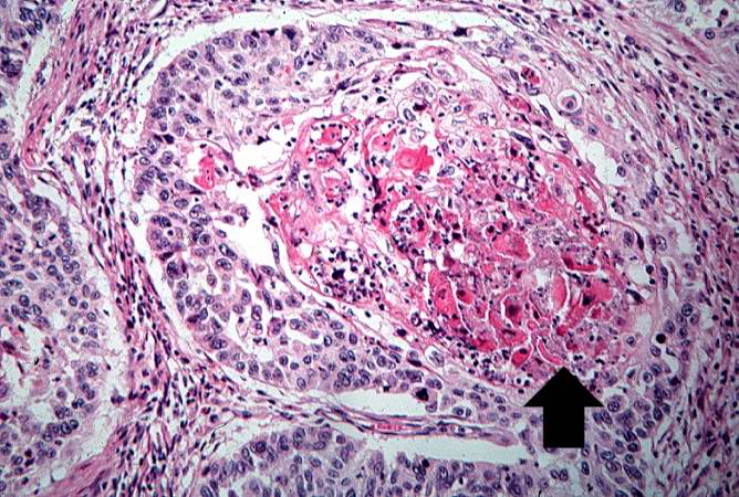
67 KB | Seung Park | This is another high-power photomicrograph showing a blood vessel with perivascular hemorrhage and mild perivascular lymphocytic cuffing (arrow). In addition, there are numerous red shrunken neurons and glia with pyknotic nuclei throughout this section. | 1 |
| 02:35, 21 August 2013 | IPLab8HSVEncephalitis5.jpg (file) |  |
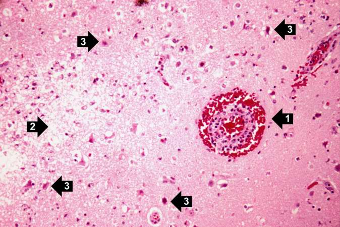
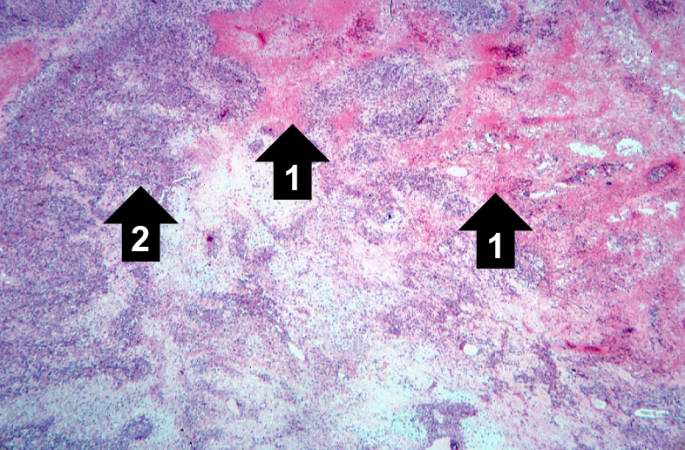
62 KB | Seung Park | This is a high-power photomicrograph of the previous section. At this power it is easier to see the blood vessel with the perivascular hemorrhage and mild perivascular lymphocytic cuffing (1). In addition, the areas of edema and loss of neurophil (2) c... | 1 |
| 02:35, 21 August 2013 | IPLab8HSVEncephalitis4.jpg (file) |  |
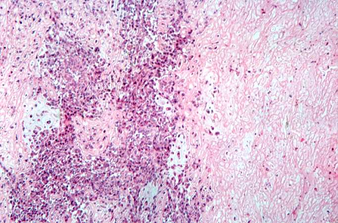
58 KB | Seung Park | This is a medium-power photomicrograph showing a blood vessel with perivascular hemorrhage (1), areas with loss of brain parenchyma, and edema (2). Even at this power, it can be seen that many of the cells are shrunken and dark red, suggesting that the... | 1 |
| 02:34, 21 August 2013 | IPLab8HSVEncephalitis3.jpg (file) |  |
41 KB | Seung Park | This is a low-power photomicrograph showing a section of brain with numerous perivascular hemorrhages (arrows) and some areas that appear hypercellular. | 1 |
| 02:34, 21 August 2013 | IPLab8HSVEncephalitis2.jpg (file) |  |
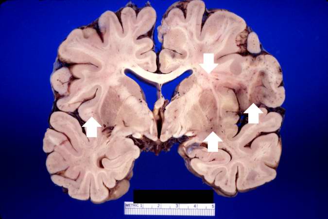
36 KB | Seung Park | This is a closer view of the previous section of brain showing multiple small, punctate hemorrhages throughout the brain parenchyma (arrows). | 1 |
| 02:34, 21 August 2013 | IPLab8HSVEncephalitis1.jpg (file) |  |
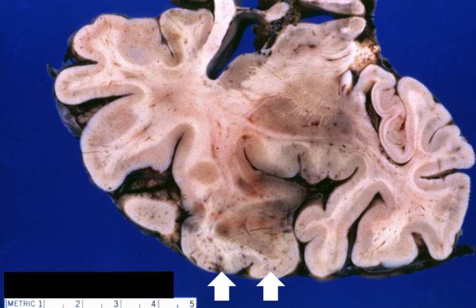
32 KB | Seung Park | This is a gross photograph of a section of brain showing multiple small, punctate hemorrhages throughout the brain parenchyma (arrows). | 1 |
| 02:29, 21 August 2013 | IPLab8HSVGlossitis5.jpg (file) |  |
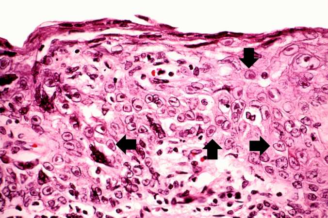
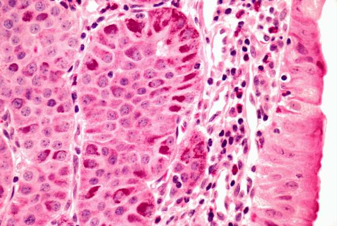
70 KB | Seung Park | This is a high-power photomicrograph of epithelium near the edge of the ulcer. The cells that have been invaded by the herpes virus contain intranuclear accumulations of amphophilic viral inclusions (arrows). | 1 |
| 02:29, 21 August 2013 | IPLab8HSVGlossitis4.jpg (file) |  |
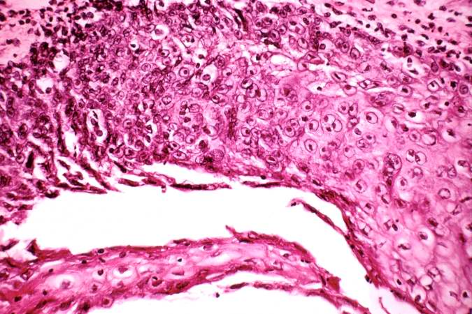
72 KB | Seung Park | This is a higher-power photomicrograph of epithelium at the edge of the ulcer. Amphophilic intranuclear inclusion bodies can be seen in almost all of the epithelial cells in this section. | 1 |
| 02:29, 21 August 2013 | IPLab8HSVGlossitis3.jpg (file) |  |
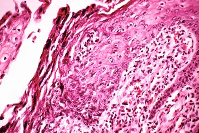
75 KB | Seung Park | This is a medium-power photomicrograph of epithelium at the edge of the ulcer. Even at this power, amphophilic (dark, blue-purple-staining) intranuclear inclusion bodies can be seen in the epithelial cells. Note the inflammatory infiltrate in the subep... | 1 |
| 02:28, 21 August 2013 | IPLab8HSVGlossitis2.jpg (file) |  |
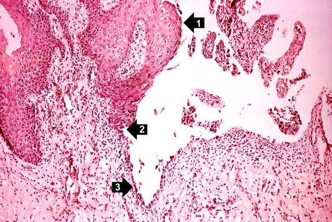
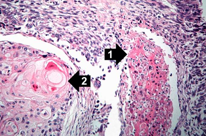
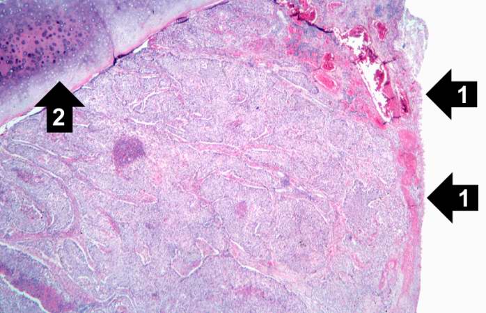
90 KB | Seung Park | This higher-power photomicrograph shows the epithelium (1), the edge of the ulcer (2), and the ulcerated epithelium (3). There is an inflammatory exudate at the base of the ulcer and some necrotic cells where the epithelium once was present. | 1 |
| 02:28, 21 August 2013 | IPLab8HSVGlossitis1.jpg (file) |  |
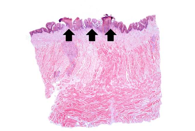
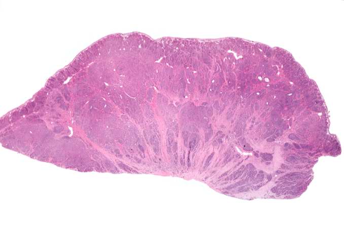
38 KB | Seung Park | This is a low-power photomicrograph showing a cross section of the tongue. There is an area along the surface of the tongue where the normal epithelium has been lost and there are areas of ulceration (arrows). | 1 |
| 02:16, 21 August 2013 | IPLab7Osteosarcoma15.jpg (file) |  |
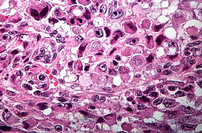
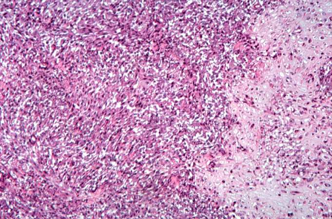
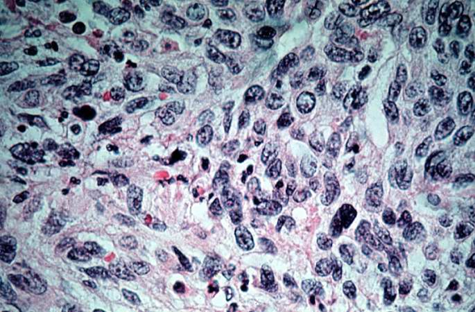
85 KB | Seung Park | This is a high-power photomicrograph of the tumor demonstrating the anaplastic cell morphology and multiple mitotic figures (arrows). | 1 |
| 02:16, 21 August 2013 | IPLab7Osteosarcoma14.jpg (file) |  |
78 KB | Seung Park | This is a high-power photomicrograph of the tumor demonstrating the anaplastic cell morphology. | 1 |
| 02:16, 21 August 2013 | IPLab7Osteosarcoma13.jpg (file) |  |
83 KB | Seung Park | This is a high-power photomicrograph of the tumor demonstrating the anaplastic cell morphology. | 1 |
| 02:15, 21 August 2013 | IPLab7Osteosarcoma12.jpg (file) |  |
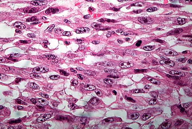
79 KB | Seung Park | This high-power photomicrograph of the tumor demonstrates the fusiform morphology of the cells. Note the marked variability in size and staining intensity of the nuclei. | 1 |
| 02:15, 21 August 2013 | IPLab7Osteosarcoma11.jpg (file) |  |
76 KB | Seung Park | This is a high-power photomicrograph of the tumor cell morphology and the periosteum (arrow). | 1 |
| 02:15, 21 August 2013 | IPLab7Osteosarcoma10.jpg (file) |  |
83 KB | Seung Park | This high-power photomicrograph demonstrates the growth pattern and the cell morphology. | 1 |
| 02:15, 21 August 2013 | IPLab7Osteosarcoma9.jpg (file) |  |
86 KB | Seung Park | This high-power photomicrograph demonstrates the cellular growth pattern. Note that the cells are fusiform and they grow in sheets. | 1 |
| 02:14, 21 August 2013 | IPLab7Osteosarcoma8.jpg (file) |  |
77 KB | Seung Park | This is a high-power photomicrograph of decalcified histologic section showing the cellularity of the tumor. | 1 |
| 02:14, 21 August 2013 | IPLab7Osteosarcoma7.jpg (file) |  |
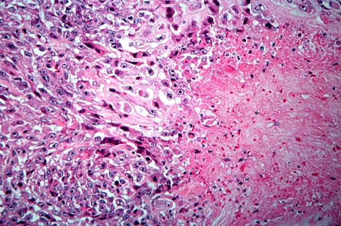
67 KB | Seung Park | This is a higher-power photomicrograph of decalcified histologic section from this tumor. There are areas of osteoid (1) and cellular areas (2). | 1 |
| 02:14, 21 August 2013 | IPLab7Osteosarcoma6.jpg (file) |  |
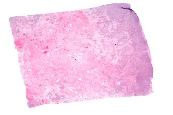
28 KB | Seung Park | This is a low-power photomicrograph of decalcified histologic section from this tumor. Note the blue color (cell nuclei stain blue) of much of this section indicating the increased cellularity of the tumor. | 1 |
| 02:14, 21 August 2013 | IPLab7Osteosarcoma5.jpg (file) |  |
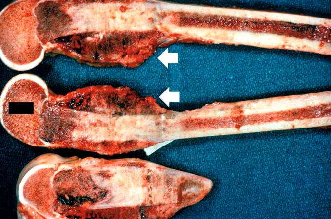
67 KB | Seung Park | These are cut sections of the distal femur containing the tumor. The periosteal involvement is evident from this picture (arrows). | 1 |
| 02:13, 21 August 2013 | IPLab7Osteosarcoma4.jpg (file) |  |
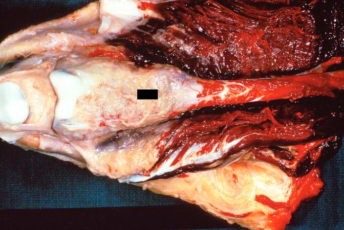
59 KB | Seung Park | This is a gross photograph of the surgical specimen with tissue dissected away to demonstrate the tumor mass. | 1 |
| 02:13, 21 August 2013 | IPLab7Osteosarcoma3.jpg (file) |  |
10 KB | Seung Park | This is another view of the tumor in the distal femur. | 1 |
| 02:13, 21 August 2013 | IPLab7Osteosarcoma2.jpg (file) |  |
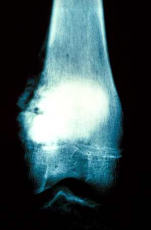
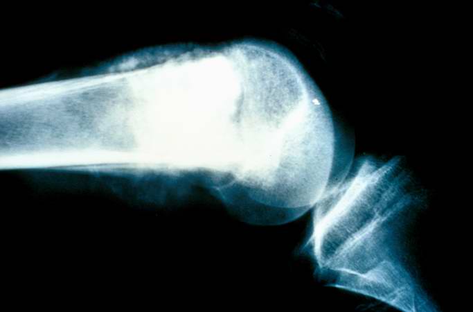
17 KB | Seung Park | This is a radiograph showing the tumor in the distal femur. | 1 |
| 02:13, 21 August 2013 | IPLab7Osteosarcoma1.jpg (file) |  |
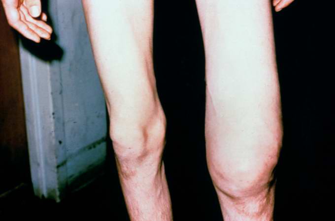
19 KB | Seung Park | This is a photograph of the patient prior to surgery. Note the marked swelling of the knee. | 1 |
| 02:08, 21 August 2013 | IPLab7Carcinoid11.jpg (file) |  |
70 KB | Seung Park | This is a high-power view of the same section stained with a silver stain to delineate carcinoid tumor cells (brown). | 1 |
| 02:08, 21 August 2013 | IPLab7Carcinoid10.jpg (file) |  |
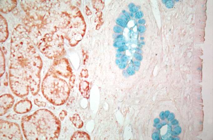
45 KB | Seung Park | This is a higher-power view of the previous section stained with a silver stain to delineate carcinoid tumor cells (brown) and a mucin stain (blue) to stain the glands. | 1 |
| 02:08, 21 August 2013 | IPLab7Carcinoid9.jpg (file) |  |
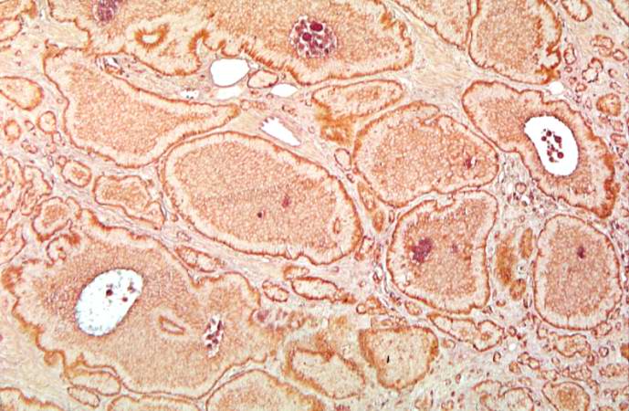
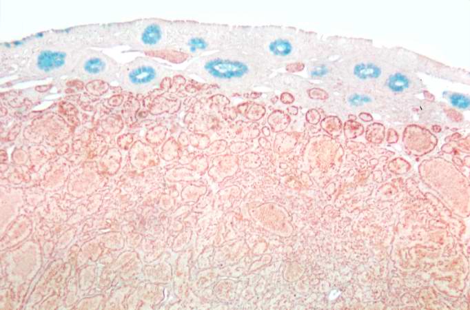
46 KB | Seung Park | This is a low-power photomicrograph of a section of cecum containing tumor stained to demonstrate the secretory granules in these tumor cells (brown-colored stain). The blue color is the mucin in the glands just under the mucosal surface. | 1 |
| 02:08, 21 August 2013 | IPLab7Carcinoid8.jpg (file) |  |
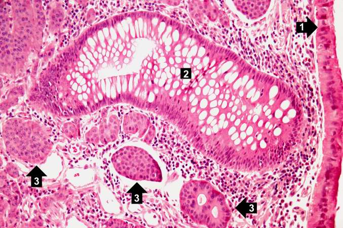
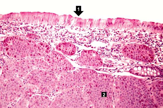
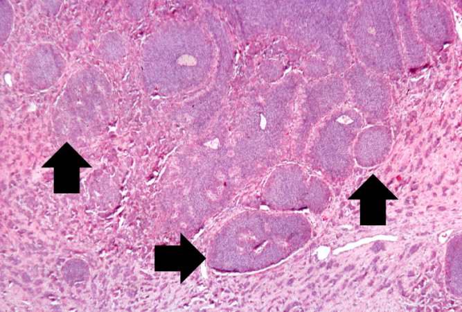
81 KB | Seung Park | This is a higher-power photomicrograph of the previous section showing intact mucosa (1), a gland (2), and the submucosal carcinoid tumor cells (3). | 1 |
| 02:07, 21 August 2013 | IPLab7Carcinoid7.jpg (file) |  |
70 KB | Seung Park | This is a low-power photomicrograph of another one of the subcutaneous masses in the cecum. The mucosa is normal and the tumor cells are in the submucosa. | 1 |
| 02:07, 21 August 2013 | IPLab7Carcinoid6.jpg (file) |  |
59 KB | Seung Park | This is a higher-power photomicrograph of the previous section showing the intact mucosa (right) and the submucosal carcinoid tumor. | 1 |
| 02:07, 21 August 2013 | IPLab7Carcinoid5.jpg (file) |  |
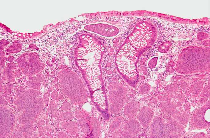
65 KB | Seung Park | This is a low-power photomicrograph of one of the subcutaneous masses in the cecum. Note that the mucosa (1) is virtually normal and the tumor cells are in the submucosa (2). | 1 |
| 02:07, 21 August 2013 | IPLab7Carcinoid4.jpg (file) |  |
77 KB | Seung Park | This is a high-power photomicrograph of the surgical specimen showing the cellular morphology. The tumor cells are monotonously similar with scant, pink, granular cytoplasm and a round-to-oval stippled nucleus. As in most carcinoid tumors, there is min... | 1 |
| 02:06, 21 August 2013 | IPLab7Carcinoid3.jpg (file) |  |
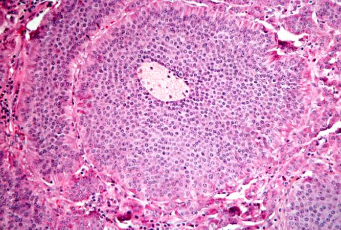
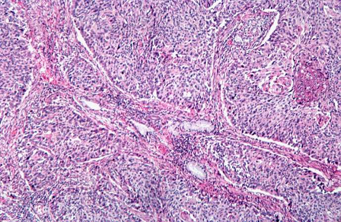
72 KB | Seung Park | This is a high-power photomicrograph of the surgical specimen showing the tumor's growth pattern--cells form discrete islands, trabeculae, and glands. | 1 |
| 02:06, 21 August 2013 | IPLab7Carcinoid2.jpg (file) |  |
62 KB | Seung Park | This is a higher-power photomicrograph of the surgical specimen showing nests of tumor cells (arrows). | 1 |
| 02:06, 21 August 2013 | IPLab7Carcinoid1.jpg (file) |  |
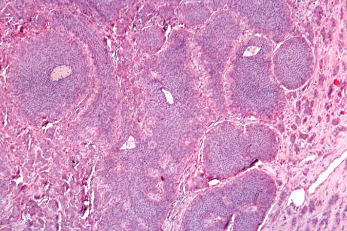
31 KB | Seung Park | This is a low-power photomicrograph of the surgical specimen showing basophilic and eosinophilic areas delimiting areas of tumor infiltration. | 1 |
| 02:01, 21 August 2013 | IPLab7Bronchogenic9.jpg (file) |  |
73 KB | Seung Park | This high-power photomicrograph of tumor shows the cytologic detail of a less-differentiated area of neoplasm with cellular anaplasia. | 1 |
| 02:01, 21 August 2013 | IPLab7Bronchogenic8.jpg (file) |  |
91 KB | Seung Park | This is a high power photomicrograph of tumor with an area of central necrosis (arrow). | 1 |
| 02:01, 21 August 2013 | IPLab7Bronchogenic7.jpg (file) |  |
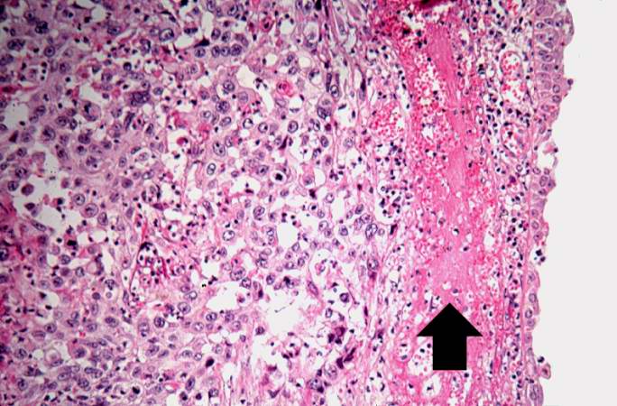
75 KB | Seung Park | This is a high-power photomicrograph showing cytologic detail of the tumor with an area of necrosis (1) and a more differentiated area with keratin pearl formation (2). | 1 |
| 02:00, 21 August 2013 | IPLab7Bronchogenic6.jpg (file) |  |
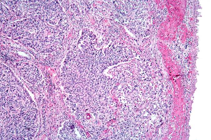
98 KB | Seung Park | This is a photomicrograph of tumor from an area of invasion with compression of fibrous stroma and focal necrosis. | 1 |
| 02:00, 21 August 2013 | IPLab7Bronchogenic5.jpg (file) |  |
71 KB | Seung Park | This is a higher-power photomicrograph of the mucosal surface (right) with an area of hemorrhage (arrow) and underlying tumor (left). | 1 |
| 02:00, 21 August 2013 | IPLab7Bronchogenic4.jpg (file) |  |
99 KB | Seung Park | This is a higher-power photomicrograph of bronchus with the ulcerated mucosal surface on the right and tumor underneath. | 1 |
| 02:00, 21 August 2013 | IPLab7Bronchogenic3.jpg (file) |  |
58 KB | Seung Park | This is a photomicrograph of bronchus with ulcerated mucosal surface on the right (1). The submucosa is completely filled with tumor down to the cartilage (2). | 1 |