IPLab:Lab 8:HSV Glossitis
Jump to navigation
Jump to search
Clinical Summary
During the course of a postmortem examination of a 58-year-old female, the tongue was noted to have a number of multiple shallow ulcers. This was an incidental finding and not related to the patient's primary disease process.
Images
-
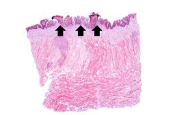 This is a low-power photomicrograph showing a cross section of the tongue. There is an area along the surface of the tongue where the normal epithelium has been lost and there are areas of ulceration (arrows).
 -
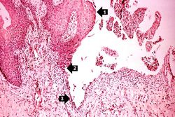 This higher-power photomicrograph shows the epithelium (1), the edge of the ulcer (2), and the ulcerated epithelium (3). There is an inflammatory exudate at the base of the ulcer and some necrotic cells where the epithelium once was present.
 -
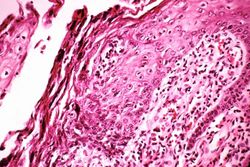 This is a medium-power photomicrograph of epithelium at the edge of the ulcer. Even at this power, amphophilic (dark, blue-purple-staining) intranuclear inclusion bodies can be seen in the epithelial cells. Note the inflammatory infiltrate in the subepithelium.
 -
 This is a higher-power photomicrograph of epithelium at the edge of the ulcer. Amphophilic intranuclear inclusion bodies can be seen in almost all of the epithelial cells in this section.
 -
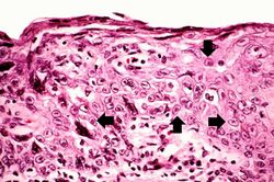 This is a high-power photomicrograph of epithelium near the edge of the ulcer. The cells that have been invaded by the herpes virus contain intranuclear accumulations of amphophilic viral inclusions (arrows).


Virtual Microscopy
Study Questions
Additional Resources
Reference
- eMedicine Medical Library: Herpes Simplex
- eMedicine Medical Library: Herpes Simplex in Emergency Medicine
- Merck Manual: Herpes Simplex Virus (HSV) Infections
Journal Articles
- Edell AR, Cohen EJ. Herpes simplex and herpes zoster eye disease: presentation and management at a city hospital for the underserved in the United States. Eye Contact Lens 2013 Jul;39(4):311-4.
Images
Related IPLab Cases
| |||||