File:IPLab5Antitrypsin6.jpg
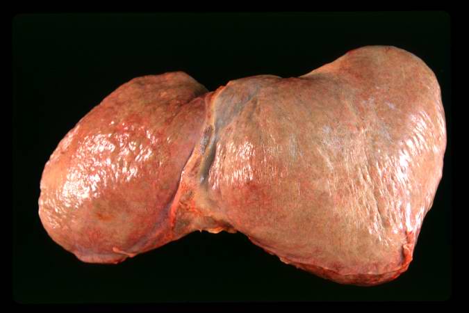
Revision as of 18:25, 19 August 2013 by Peter Anderson (talk | contribs) (This is a gross photograph of the liver from this case. The capsule is somewhat thickened and the surface is slightly roughened, though it is difficult to appreciate the nodularity of the liver.)
No higher resolution available.
IPLab5Antitrypsin6.jpg (675 × 450 pixels, file size: 29 KB, MIME type: image/jpeg)
This is a gross photograph of the liver from this case. The capsule is somewhat thickened and the surface is slightly roughened, though it is difficult to appreciate the nodularity of the liver.
File history
Click on a date/time to view the file as it appeared at that time.
| Date/Time | Thumbnail | Dimensions | User | Comment | |
|---|---|---|---|---|---|
| current | 18:25, 19 August 2013 | | 675 × 450 (29 KB) | Peter Anderson (talk | contribs) | This is a gross photograph of the liver from this case. The capsule is somewhat thickened and the surface is slightly roughened, though it is difficult to appreciate the nodularity of the liver. |
- You cannot overwrite this file.
File usage
The following page links to this file: