File:IPLab6RA6.jpg
Revision as of 18:07, 19 August 2013 by Seung Park (talk | contribs) (This higher-power photomicrograph of the subcutaneous nodule shows a granulomatous lesion with a necrotic center and a peripheral rim of macrophages, fibrocytes, and occasional lymphocytes. In the necrotic center of the granuloma there is some minerali...)
No higher resolution available.
IPLab6RA6.jpg (677 × 450 pixels, file size: 64 KB, MIME type: image/jpeg)
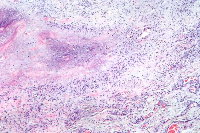
This higher-power photomicrograph of the subcutaneous nodule shows a granulomatous lesion with a necrotic center and a peripheral rim of macrophages, fibrocytes, and occasional lymphocytes. In the necrotic center of the granuloma there is some mineralization (basophilic material).
File history
Click on a date/time to view the file as it appeared at that time.
| Date/Time | Thumbnail | Dimensions | User | Comment | |
|---|---|---|---|---|---|
| current | 18:07, 19 August 2013 | | 677 × 450 (64 KB) | Seung Park (talk | contribs) | This higher-power photomicrograph of the subcutaneous nodule shows a granulomatous lesion with a necrotic center and a peripheral rim of macrophages, fibrocytes, and occasional lymphocytes. In the necrotic center of the granuloma there is some minerali... |
- You cannot overwrite this file.
File usage
There are no pages that link to this file.