File:IPLab2Calcification7.jpg
No higher resolution available.
IPLab2Calcification7.jpg (680 × 450 pixels, file size: 59 KB, MIME type: image/jpeg)
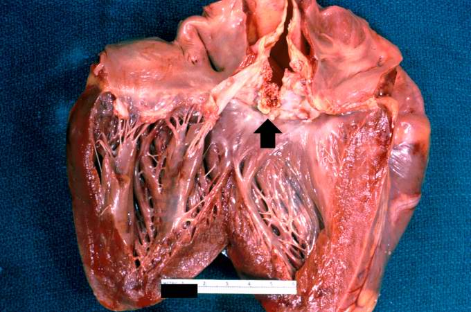
A closer view of this same aortic valve (arrow) illustrates the nodularity and thickening of this valve. This valve would be extremely stiff and almost entirely immobile. This particular example of dystrophic calcification is associated with a degenerative change of the aortic valve due to an unknown cause.
File history
Click on a date/time to view the file as it appeared at that time.
| Date/Time | Thumbnail | Dimensions | User | Comment | |
|---|---|---|---|---|---|
| current | 11:36, 19 August 2013 | | 680 × 450 (59 KB) | Peter Anderson (talk | contribs) | A closer view of this same aortic valve (arrow) illustrates the nodularity and thickening of this valve. This valve would be extremely stiff and almost entirely immobile. This particular example of dystrophic calcification is associated with a degenera... |
You cannot overwrite this file.
File usage
The following page uses this file:
