From Pathology Education Instructional Resource
Jump to navigation
Jump to search
Images
-
This is a gross photograph of the heart from this case demonstrating the well-formed
thrombus (arrow) tightly attached to the myocardium near the apex of the left ventricle.
-
This is a low-power photomicrograph of the
thrombus (1) attached to the myocardium (2).
-
This higher-power photomicrograph shows the border between the
thrombus on the right (1) and the endocardium on the left (2). There is a line of inflammatory cells at this interface (arrow).
-
This is a high-power photomicrograph of the border zone between the
thrombus (1) and the endocardium (2). In this region there is less inflammation at the border zone.
-
This photomicrograph illustrates the layered effect of the
thrombus.
-
This is a higher-power photomicrograph of the
thrombus. Note the pale regions which contain primarily platelets (degranulated platelets) with some fibrin (1), and the red areas which contain RBCs, some leukocytes, and fibrin(2).
-
This high-power photomicrograph of
thrombus demonstrates more clearly the components of the layers--the pale regions which contain primarily platelets (degranulated platelets) with some fibrin (1), and the red areas which contain RBCs, some leukocytes, and fibrin (2).
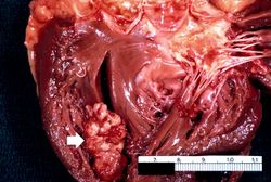 This is a gross photograph of the heart from this case demonstrating the well-formed thrombus (arrow) tightly attached to the myocardium near the apex of the left ventricle.
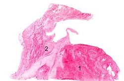 This is a low-power photomicrograph of the thrombus (1) attached to the myocardium (2).
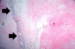 This higher-power photomicrograph shows the border between the thrombus on the right (1) and the endocardium on the left (2). There is a line of inflammatory cells at this interface (arrow).
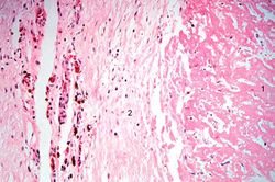 This is a high-power photomicrograph of the border zone between the thrombus (1) and the endocardium (2). In this region there is less inflammation at the border zone.
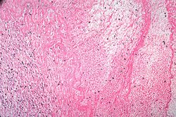 This photomicrograph illustrates the layered effect of the thrombus.
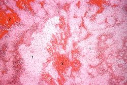 This is a higher-power photomicrograph of the thrombus. Note the pale regions which contain primarily platelets (degranulated platelets) with some fibrin (1), and the red areas which contain RBCs, some leukocytes, and fibrin(2).
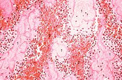 This high-power photomicrograph of thrombus demonstrates more clearly the components of the layers--the pale regions which contain primarily platelets (degranulated platelets) with some fibrin (1), and the red areas which contain RBCs, some leukocytes, and fibrin (2).


