File:IPLab3ChronicPepticUlcer11.jpg
Revision as of 04:17, 19 August 2013 by Seung Park (talk | contribs) (This is a trichrome-stained section of tissue demonstrating fibrous connective tissue scar formation (blue color) in this lesion. The surface of the ulcer is at the left-hand side of the image. There is a layer of inflammatory cells and RBCs on the sur...)
No higher resolution available.
IPLab3ChronicPepticUlcer11.jpg (683 × 450 pixels, file size: 73 KB, MIME type: image/jpeg)
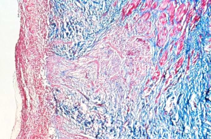
This is a trichrome-stained section of tissue demonstrating fibrous connective tissue scar formation (blue color) in this lesion. The surface of the ulcer is at the left-hand side of the image. There is a layer of inflammatory cells and RBCs on the surface of the ulcer.
File history
Click on a date/time to view the file as it appeared at that time.
| Date/Time | Thumbnail | Dimensions | User | Comment | |
|---|---|---|---|---|---|
| current | 04:17, 19 August 2013 | | 683 × 450 (73 KB) | Seung Park (talk | contribs) | This is a trichrome-stained section of tissue demonstrating fibrous connective tissue scar formation (blue color) in this lesion. The surface of the ulcer is at the left-hand side of the image. There is a layer of inflammatory cells and RBCs on the sur... |
- You cannot overwrite this file.
File usage
There are no pages that link to this file.