File:IPLab3Sarcoidosis4.jpg
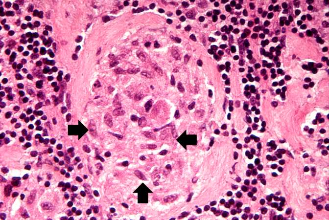
Revision as of 03:30, 19 August 2013 by Seung Park (talk | contribs) (This photomicrograph of a single granuloma illustrates the individual macrophages (arrows) which make up the bulk of this tissue. There is an absence of necrosis in the center of the lesions in this case.)
No higher resolution available.
IPLab3Sarcoidosis4.jpg (672 × 450 pixels, file size: 74 KB, MIME type: image/jpeg)
This photomicrograph of a single granuloma illustrates the individual macrophages (arrows) which make up the bulk of this tissue. There is an absence of necrosis in the center of the lesions in this case.
File history
Click on a date/time to view the file as it appeared at that time.
| Date/Time | Thumbnail | Dimensions | User | Comment | |
|---|---|---|---|---|---|
| current | 03:30, 19 August 2013 | | 672 × 450 (74 KB) | Seung Park (talk | contribs) | This photomicrograph of a single granuloma illustrates the individual macrophages (arrows) which make up the bulk of this tissue. There is an absence of necrosis in the center of the lesions in this case. |
- You cannot overwrite this file.
File usage
The following page links to this file: