File:IPLab12Mesothelioma4.jpg
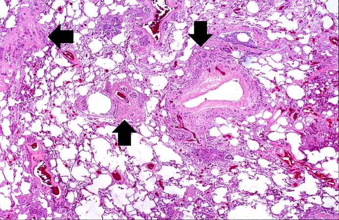
Revision as of 05:33, 21 August 2013 by Seung Park (talk | contribs) (This is a low-power photomicrograph of a section of the left lung. At this magnification you can see areas of consolidation (arrows), especially around blood vessels.)
No higher resolution available.
IPLab12Mesothelioma4.jpg (693 × 450 pixels, file size: 113 KB, MIME type: image/jpeg)
This is a low-power photomicrograph of a section of the left lung. At this magnification you can see areas of consolidation (arrows), especially around blood vessels.
File history
Click on a date/time to view the file as it appeared at that time.
| Date/Time | Thumbnail | Dimensions | User | Comment | |
|---|---|---|---|---|---|
| current | 05:33, 21 August 2013 | | 693 × 450 (113 KB) | Seung Park (talk | contribs) | This is a low-power photomicrograph of a section of the left lung. At this magnification you can see areas of consolidation (arrows), especially around blood vessels. |
- You cannot overwrite this file.
File usage
The following page links to this file: