File:IPLab10Histo7.jpg
Revision as of 04:07, 21 August 2013 by Seung Park (talk | contribs) (This photomicrograph was taken under oil immersion to show the silver-stained Histoplasma organisms. Some of the organisms appear to be budding (arrows).)
No higher resolution available.
IPLab10Histo7.jpg (668 × 450 pixels, file size: 34 KB, MIME type: image/jpeg)
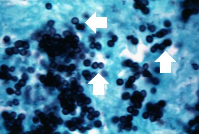
This photomicrograph was taken under oil immersion to show the silver-stained Histoplasma organisms. Some of the organisms appear to be budding (arrows).
File history
Click on a date/time to view the file as it appeared at that time.
| Date/Time | Thumbnail | Dimensions | User | Comment | |
|---|---|---|---|---|---|
| current | 04:07, 21 August 2013 | | 668 × 450 (34 KB) | Seung Park (talk | contribs) | This photomicrograph was taken under oil immersion to show the silver-stained Histoplasma organisms. Some of the organisms appear to be budding (arrows). |
- You cannot overwrite this file.
File usage
The following page links to this file: