File list
This special page shows all uploaded files.
| Date | Name | Thumbnail | Size | User | Description | Versions |
|---|---|---|---|---|---|---|
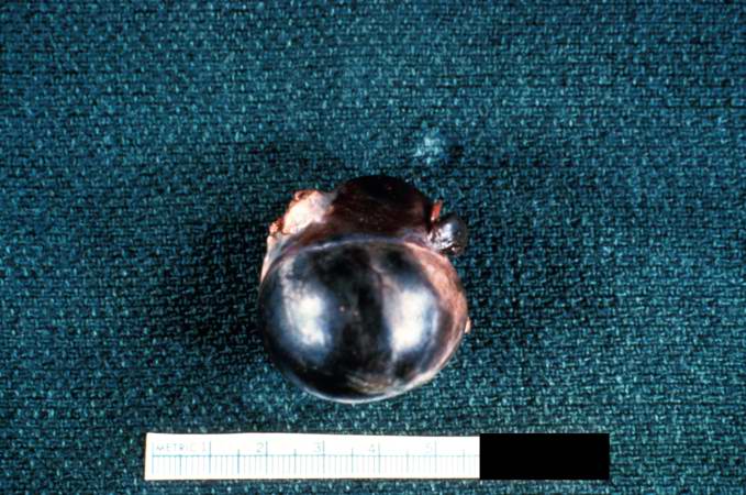
| 16:48, 19 August 2013 | IPLab4Thromboembolus7.jpg (file) |  |
64 KB | Seung Park | This is a gross photograph of an infarcted testis. Because of the anatomy of the blood supply to the testis, torsion or the blood vessels often leads to venous occlusion (due to compression of the thin walled veins) but not arterial occlusion. Thus, bl... | 1 |
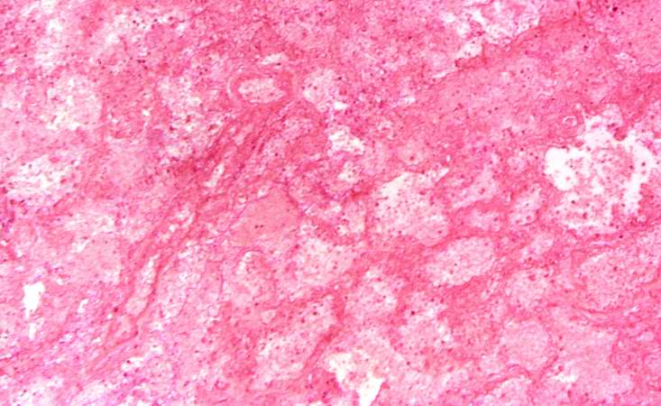
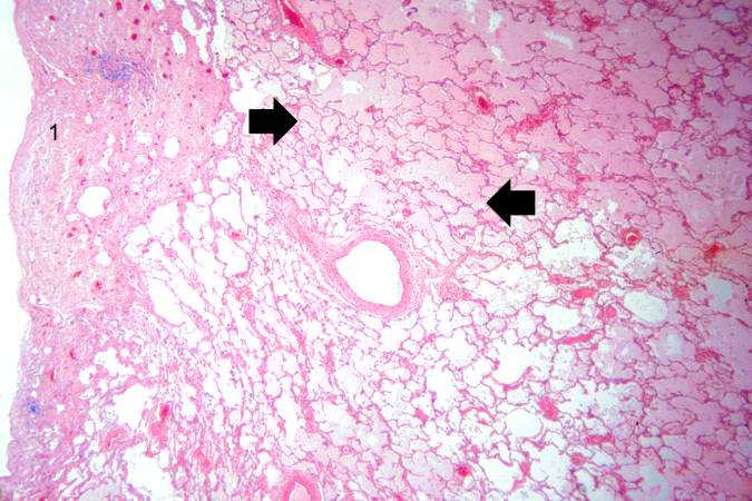
| 16:48, 19 August 2013 | IPLab4Thromboembolus6.jpg (file) |  |
61 KB | Seung Park | This is a low-power photomicrograph of the infarcted lung. The tissue is congested and has a very bland appearance due to coagulation necrosis of the lung parenchyma. You can still see the outlines of the alveoli and the cells that make-up the alveoli ... | 1 |
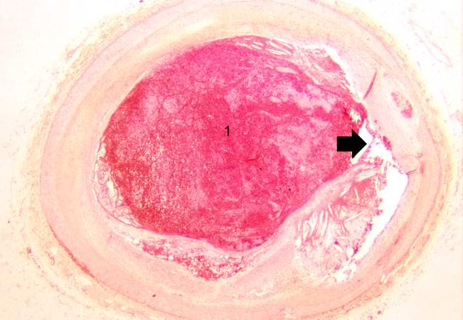
| 16:48, 19 August 2013 | IPLab4Thromboembolus5.jpg (file) |  |
66 KB | Seung Park | This is a photomicrograph of the wall of the pulmonary artery (1) containing the thromboembolus. In this case the artery wall looks normal. If this was a thrombus instead of a thromboembolus, you would expect to see some damage in the artery wall that ... | 1 |
| 16:47, 19 August 2013 | IPLab4Thromboembolus4.jpg (file) |  |
47 KB | Seung Park | This is a low-power photomicrograph of lung. A large thrombus is lodged at this branch point in the pulmonary artery. Note the hemorrhage and congestion in the surrounding lung parenchyma. | 1 |
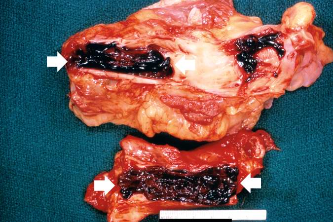
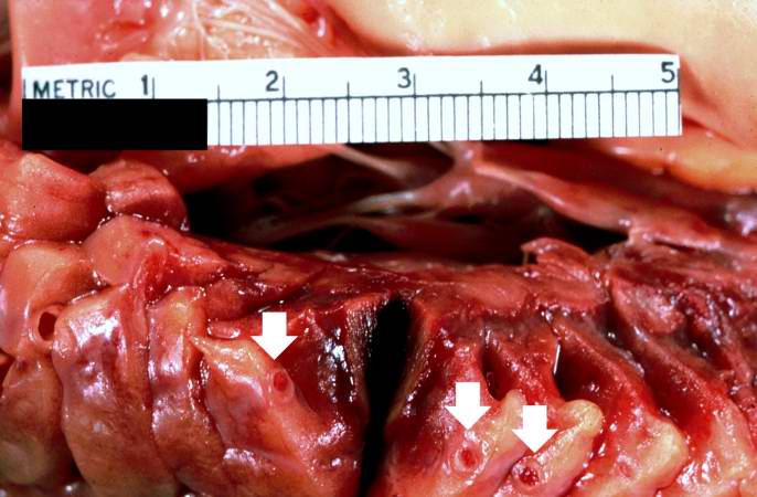
| 16:47, 19 August 2013 | IPLab4Thromboembolus3.jpg (file) |  |
64 KB | Seung Park | This is a gross photograph of portions of muscle from the legs including sections of leg veins. Note that the leg veins contain thrombus (arrows). | 1 |
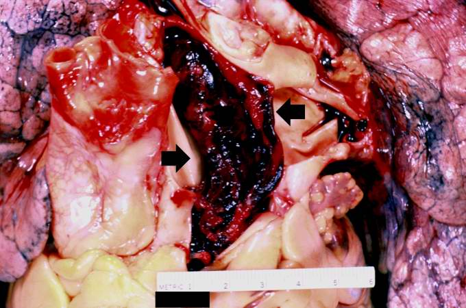
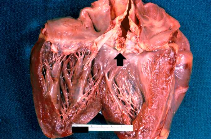
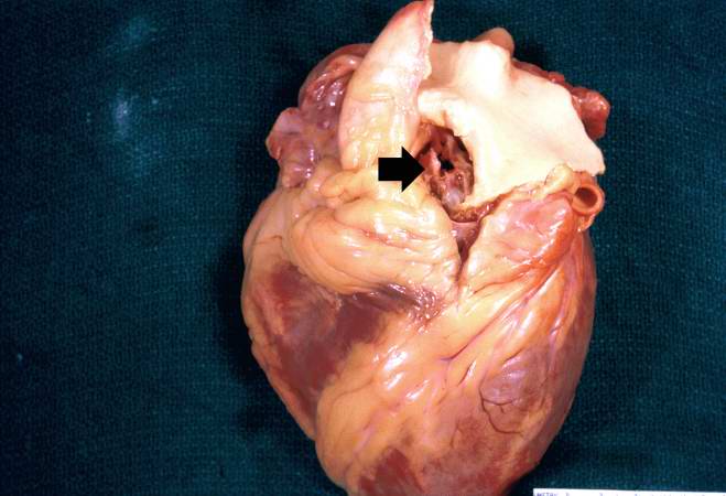
| 16:46, 19 August 2013 | IPLab4Thromboembolus2.jpg (file) |  |
57 KB | Seung Park | This is a gross photograph of the heart with the main pulmonary artery opened. Note the thromboembolus filling the pulmonary artery (arrows). | 1 |
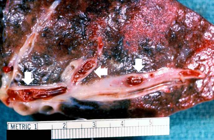
| 16:46, 19 August 2013 | IPLab4Thromboembolus1.jpg (file) |  |
63 KB | Seung Park | This is a gross photograph of a cut section of lung demonstrating thromboemboli in the pulmonary arteries (arrows). | 1 |
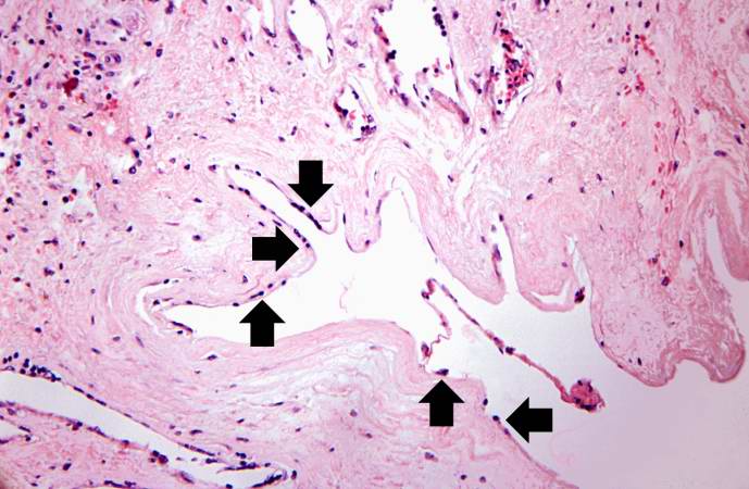
| 16:41, 19 August 2013 | IPLab4Thrombosis10.jpg (file) |  |
54 KB | Seung Park | This is a high-power photomicrograph of the luminal surface of a re-canalized vessel. Note that the vessel lumen is lined by endothelial cells (arrows). | 1 |
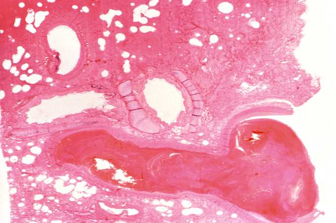
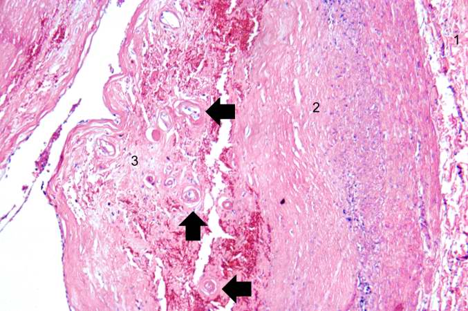
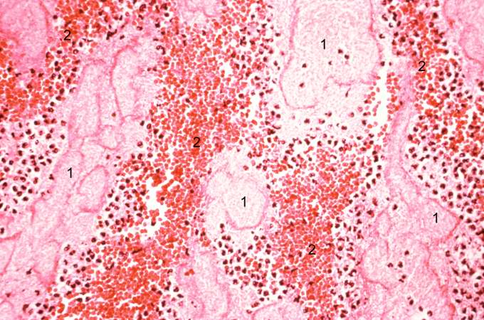
| 16:40, 19 August 2013 | IPLab4Thrombosis9.jpg (file) |  |
67 KB | Seung Park | This is a higher-power photomicrograph of another region of the vessel wall. The adventitia (1) and the media (2) contain inflammatory cells. The recanalized portion of the vessel (3) is composed of fibrous connective tissue and contains numerous small... | 1 |
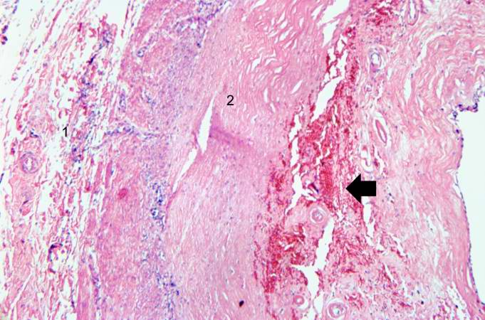
| 16:40, 19 August 2013 | IPLab4Thrombosis8.jpg (file) |  |
65 KB | Seung Park | This is a higher-power photomicrograph of the vessel wall. The adventitia (1) and the media (2) contain inflammatory cells. The recanalized portion of the vessel is composed of fibrous connective tissue and contains numerous small blood vessels. There ... | 1 |
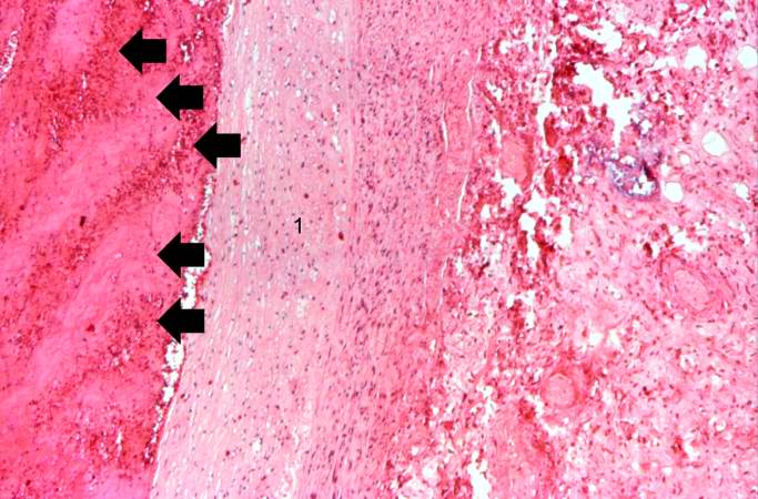
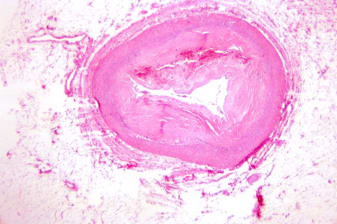
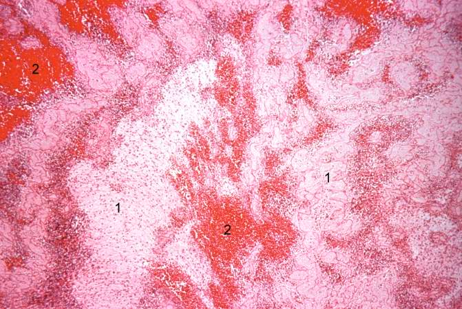
| 16:40, 19 August 2013 | IPLab4Thrombosis7.jpg (file) |  |
50 KB | Seung Park | In this low-power photomicrograph of another coronary artery from this patient, a mural thrombus has undergone re-organization. The mural thrombus has been invaded by the in-growth of fibroblasts and small blood vessels from the wall of the artery. The... | 1 |
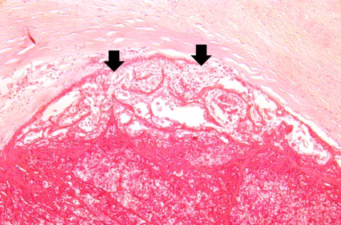
| 16:39, 19 August 2013 | IPLab4Thrombosis6.jpg (file) |  |
66 KB | Seung Park | This is a higher-power photomicrograph of thrombus attached to the wall of the vessel. Note the early organization with in-growth of fibroblasts and small blood vessels from the wall of the artery (arrows). | 1 |
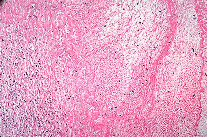
| 16:38, 19 August 2013 | IPLab4Thrombosis5.jpg (file) |  |
57 KB | Seung Park | This is a high-power photomicrograph of thrombus attached to the wall of the vessel. There is early organization of the thrombus (arrow). | 1 |
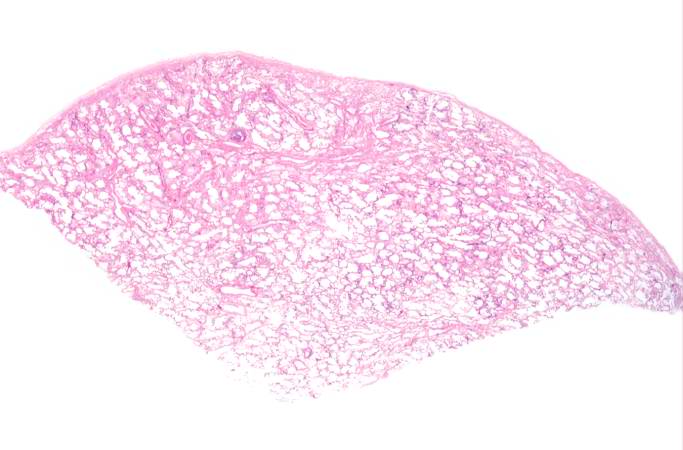
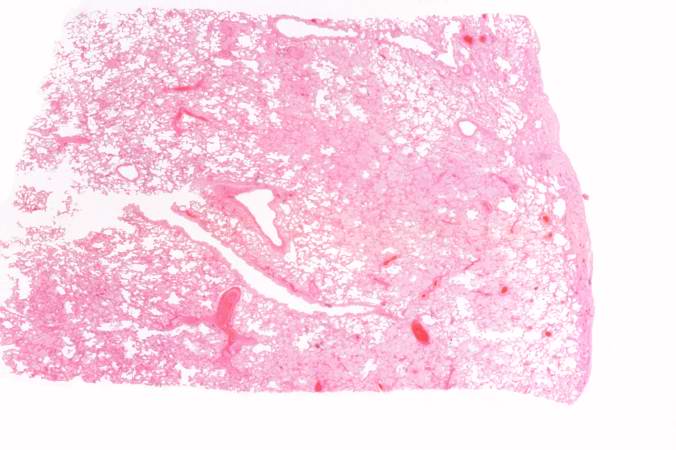
| 16:38, 19 August 2013 | IPLab2Calcification2.jpg (file) |  |
37 KB | Peter Anderson | This low-power photomicrograph of the patient's lung illustrates large, open alveolar spaces. The pleural surface is the curved surface at the top. | 1 |
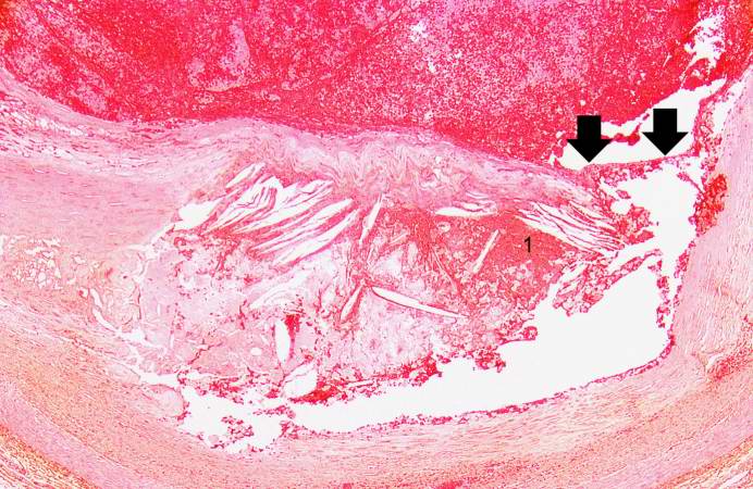
| 16:36, 19 August 2013 | IPLab4Thrombosis4.jpg (file) |  |
65 KB | Seung Park | This is another high-power photomicrograph of the ruptured fibrous cap (arrows) with hemorrhage (1) into the atherosclerotic plaque. Note the presence of cholesterol crystals. | 1 |
| 16:36, 19 August 2013 | IPLab2Calcification8.jpg (file) |  |
45 KB | Peter Anderson | This gross photograph affords a closer view of the same aortic valve. Note the nodularity and thickening of this valve due to fibrosis and dystrophic calcification. | 1 |
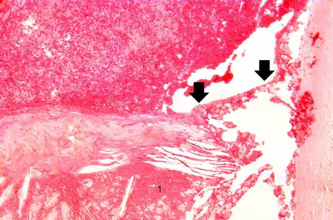
| 16:36, 19 August 2013 | IPLab4Thrombosis3.jpg (file) |  |
81 KB | Seung Park | This is a higher-power photomicrograph of the ruptured fibrous cap (arrows) with hemorrhage (1) into the atherosclerotic plaque. | 1 |
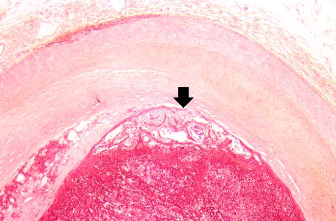
| 16:36, 19 August 2013 | IPLab2Calcification7.jpg (file) |  |
59 KB | Peter Anderson | A closer view of this same aortic valve (arrow) illustrates the nodularity and thickening of this valve. This valve would be extremely stiff and almost entirely immobile. This particular example of dystrophic calcification is associated with a degenera... | 1 |
| 16:35, 19 August 2013 | IPLab4Thrombosis2.jpg (file) |  |
42 KB | Seung Park | This is a low-power photomicrograph of thrombosed coronary artery. The thrombus (1) completely occludes the vessel. Note the layering of the thrombus. The fibrous cap is ruptured (arrow) and there is hemorrhage into the atherosclerotic plaque. Note the... | 1 |
| 16:35, 19 August 2013 | IPLab2Calcification6.jpg (file) |  |
39 KB | Peter Anderson | Metastatic calcification is only one of two forms of pathologic calcification. Unlike metastatic calcification, dystrophic calcification does not require an increase in serum calcium levels. This is a gross specimen of a heart with dystrophic calcifica... | 1 |
| 16:35, 19 August 2013 | IPLab4Thrombosis1.jpg (file) |  |
47 KB | Seung Park | This is a gross photograph of thrombosed coronary artery (arrows). | 1 |
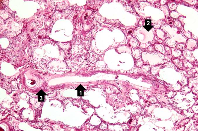
| 16:34, 19 August 2013 | IPLab2Calcification5.jpg (file) |  |
68 KB | Peter Anderson | This photomicrograph demonstrates pulmonary alveoli with extensive calcium depositions (1) in the septa and protein accumulations (2) in the alveoli. | 1 |
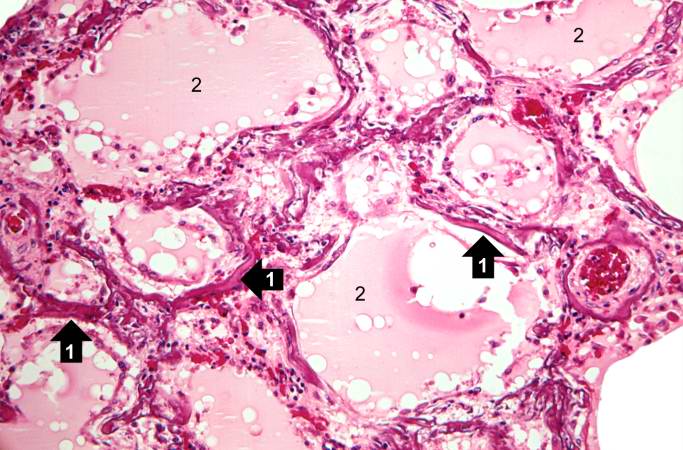
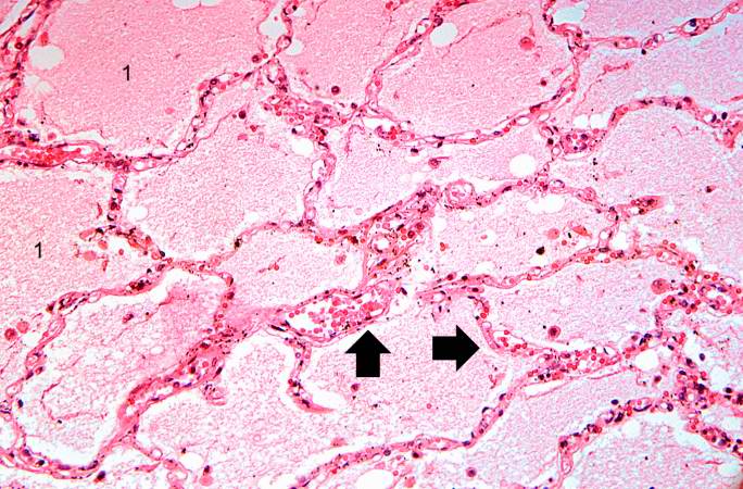
| 16:34, 19 August 2013 | IPLab2Calcification4.jpg (file) |  |
65 KB | Peter Anderson | This high-power photomicrograph of a blood vessel shows calcium deposits in the vascular wall (1) and proteinaceous material (2) (from edema) within some of the alveoli. The smooth muscle in the vessel wall has been almost completely replaced by calciu... | 1 |
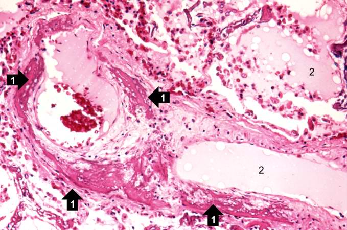
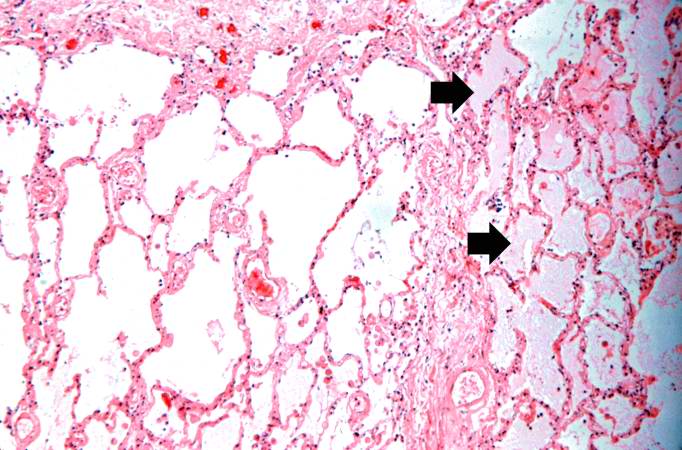
| 16:33, 19 August 2013 | IPLab2Calcification3.jpg (file) |  |
78 KB | Peter Anderson | A higher-power photomicrograph shows a blood vessel cut in longitudinal section (1). Several of the alveoli are filled with a pink-staining proteinaceous fluid (2) indicative of pulmonary edema. The alveolar septa and the wall of the blood vessel have ... | 1 |
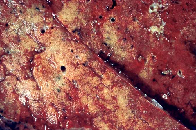
| 16:32, 19 August 2013 | IPLab2Calcification1.jpg (file) |  |
72 KB | Peter Anderson | This is a gross photograph of the cut section of the patient's lung showing evidence of severe metastatic calcification. The lung tissue has a rough, firm appearance with open airways. | 1 |
| 16:30, 19 August 2013 | IPLab4MuralThrombus7.jpg (file) |  |
80 KB | Seung Park | This high-power photomicrograph of thrombus demonstrates more clearly the components of the layers--the pale regions which contain primarily platelets (degranulated platelets) with some fibrin (1), and the red areas which contain RBCs, some leukocytes,... | 1 |
| 16:29, 19 August 2013 | IPLab4MuralThrombus6.jpg (file) |  |
82 KB | Seung Park | This is a higher-power photomicrograph of the thrombus. Note the pale regions which contain primarily platelets (degranulated platelets) with some fibrin (1), and the red areas which contain RBCs, some leukocytes, and fibrin(2). | 1 |
| 16:29, 19 August 2013 | IPLab4MuralThrombus5.jpg (file) |  |
93 KB | Seung Park | This photomicrograph illustrates the layered effect of the thrombus. | 1 |
| 16:28, 19 August 2013 | IPLab4MuralThrombus4.jpg (file) |  |
65 KB | Seung Park | This is a high-power photomicrograph of the border zone between the thrombus (1) and the endocardium (2). In this region there is less inflammation at the border zone. | 1 |
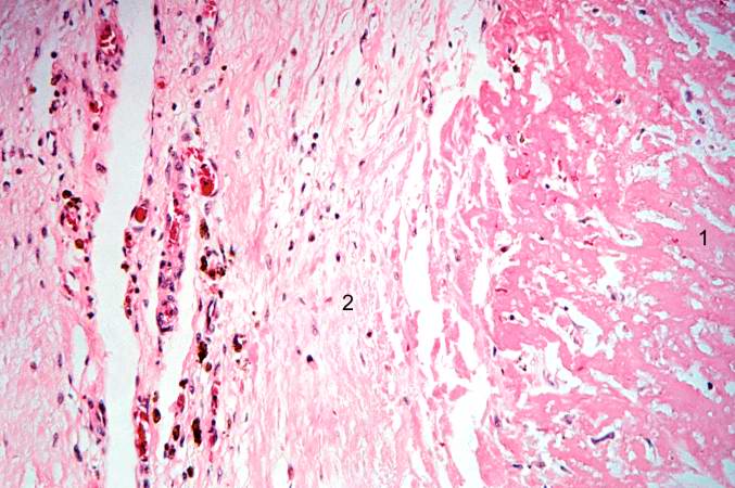
| 16:28, 19 August 2013 | IPLab4MuralThrombus3.jpg (file) |  |
53 KB | Seung Park | This higher-power photomicrograph shows the border between the thrombus on the right (1) and the endocardium on the left (2). There is a line of inflammatory cells at this interface (arrow). | 1 |
| 16:27, 19 August 2013 | IPLab4MuralThrombus2.jpg (file) |  |
39 KB | Seung Park | This is a low-power photomicrograph of the thrombus (1) attached to the myocardium (2). | 1 |
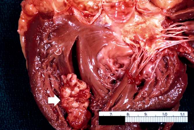
| 16:27, 19 August 2013 | IPLab4MuralThrombus1.jpg (file) |  |
60 KB | Seung Park | This is a gross photograph of the heart from this case demonstrating the well-formed thrombus (arrow) tightly attached to the myocardium near the apex of the left ventricle. | 1 |
| 16:22, 19 August 2013 | IPLab4ChronicPassiveCongestion9.jpg (file) |  |
59 KB | Seung Park | This is a gross photograph of the cut surface of a liver with chronic passive congestion (left) compared to the cut surface of a nutmeg (right). | 1 |
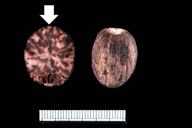
| 16:22, 19 August 2013 | IPLab4ChronicPassiveCongestion8.jpg (file) |  |
21 KB | Seung Park | This is a gross photograph of a nutmeg. You can see from the appearance of the cut surface of the nutmeg (arrow) why chronic passive congestion of the liver is sometimes referred to as "nutmeg liver." | 1 |
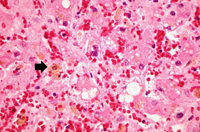
| 16:21, 19 August 2013 | IPLab4ChronicPassiveCongestion7.jpg (file) |  |
61 KB | Seung Park | This is a high-power photomicrograph of liver with several macrophages that are distended with a brown pigment (arrow). These resident macrophages (Kupffer cells) are part of the reticuloendothelial system and normally line the sinusoidal spaces in the... | 1 |
| 16:21, 19 August 2013 | IPLab4ChronicPassiveCongestion6.jpg (file) |  |
76 KB | Seung Park | This is a high-power photomicrograph of the central vein illustrating congestion and some loss of liver parenchymal cells. A mild increase in connective tissue around the central vein is evident in this section. | 1 |
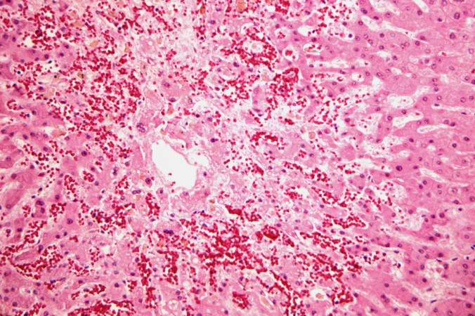
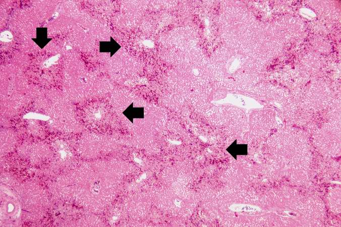
| 16:20, 19 August 2013 | IPLab4ChronicPassiveCongestion5.jpg (file) |  |
83 KB | Seung Park | This higher-power photomicrograph of the liver lobules shows congestion and red blood cell accumulation in the sinusoidal spaces around the central vein. Note that around the portal triads (arrows) the liver cells are quite normal and there is no evide... | 1 |
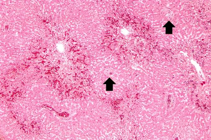
| 16:20, 19 August 2013 | IPLab4ChronicPassiveCongestion4.jpg (file) |  |
83 KB | Seung Park | This is a higher-power photomicrograph of liver demonstrating an accentuated lobular pattern with a dark red stain surrounding the central veins in the liver lobules (arrows). | 1 |
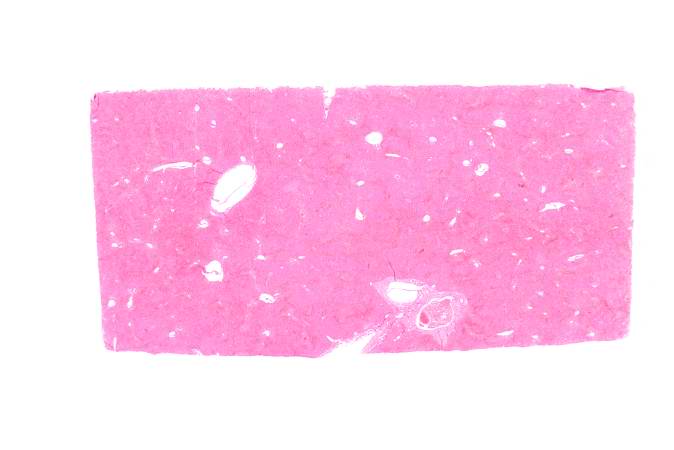
| 16:20, 19 August 2013 | IPLab4ChronicPassiveCongestion3.jpg (file) |  |
21 KB | Seung Park | This low-power photomicrograph of liver demonstrates a slightly visible pattern of centrilobular congestion at this magnification. | 1 |
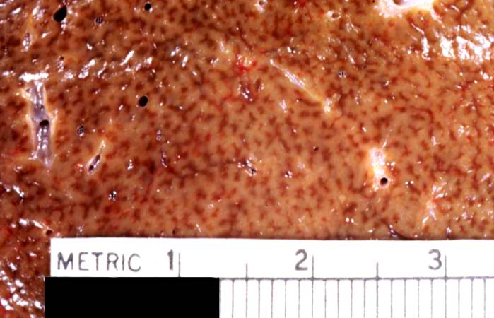
| 16:19, 19 August 2013 | IPLab4ChronicPassiveCongestion2.jpg (file) |  |
44 KB | Seung Park | This is a closer view of a cut section of liver demonstrating the pattern of chronic passive congestion. The central vein regions are red and the surrounding hepatic tissue is pale tan-brown. | 1 |
| 16:19, 19 August 2013 | IPLab4ChronicPassiveCongestion1.jpg (file) |  |
43 KB | Seung Park | This is a gross photograph of a liver demonstrating chronic passive congestion. Note the accentuation of the centrilobular pattern evidenced by the dark-brown-staining areas in this tissue. | 1 |
| 16:12, 19 August 2013 | IPLab4PulmonaryCongestion7.jpg (file) |  |
82 KB | Seung Park | This high-power photomicrograph illustrates the edema fluid within the alveoli (1) and the congestion (RBCs) in the alveolar capillaries (arrows). | 1 |
| 16:12, 19 August 2013 | IPLab4PulmonaryCongestion6.jpg (file) |  |
65 KB | Seung Park | This is a higher-power photomicrograph showing edema-filled alveoli in the right portion of this section (arrows). | 1 |
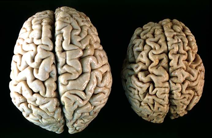
| 16:12, 19 August 2013 | IPLab2Atrophy10.jpg (file) |  |
43 KB | Peter Anderson | This gross photograph shows a normal brain (left) and a brain from a geriatric patient (right). Note the decreased size, the narrowed gyri, and the widened sulci of the brain from this octogenarian. What is the cause of atrophy in this case? | 1 |
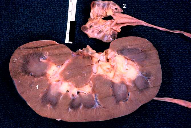
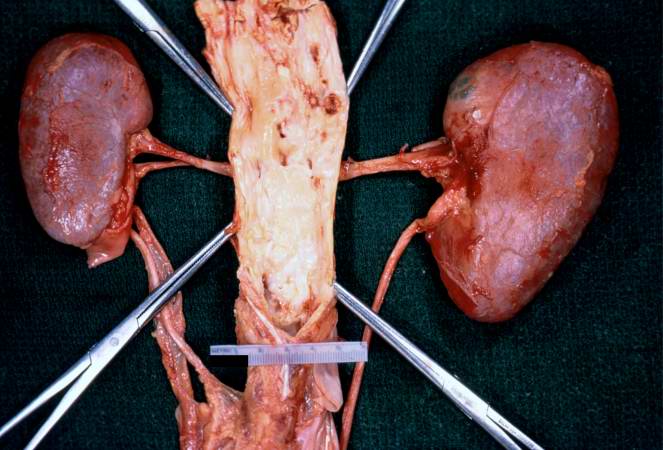
| 16:11, 19 August 2013 | IPLab2Atrophy9.jpg (file) |  |
47 KB | Peter Anderson | The two kidneys in this slide are from the same patient. One kidney (1) is relatively normal, although increased in size due to compensatory hypertrophy. The other kidney (2) is very small with only rudimentary nodules of renal parenchyma. This kidney ... | 1 |
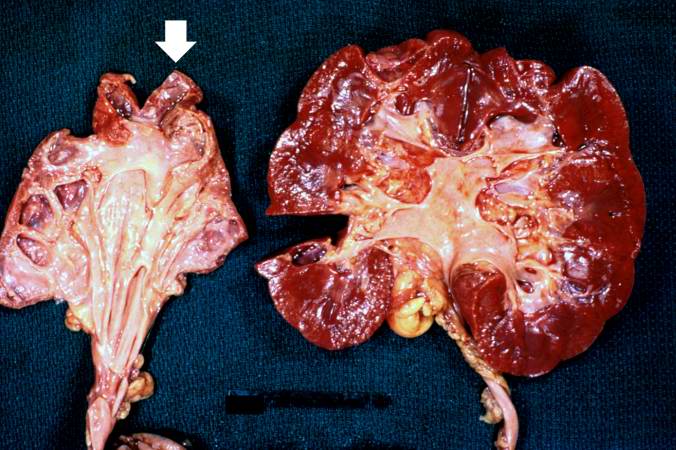
| 16:11, 19 August 2013 | IPLab2Atrophy8.jpg (file) |  |
64 KB | Peter Anderson | These kidneys were removed from a patient who had blockage of one ureter leading to increased pressure in the renal pelvis. The increased pressure produced hydronephrosis (arrow) in one kidney. What is the cause of atrophy in this case? | 1 |
| 16:10, 19 August 2013 | IPLab2Atrophy7.jpg (file) |  |
50 KB | Peter Anderson | In this gross photograph of kidneys and the abdominal aorta, there is narrowing of the left renal artery at its ostium from the aorta. This atherosclerotic narrowing of the renal artery causes reduced blood pressure in the kidney whose artery is affect... | 1 |
| 16:08, 19 August 2013 | IPLab4PulmonaryCongestion5.jpg (file) |  |
63 KB | Seung Park | This is a higher-power photomicrograph of lung. The edema fluid within the alveoli is visible at this higher magnification (arrows). The thickened pleura (1) is on the left. | 1 |
| 16:07, 19 August 2013 | IPLab4PulmonaryCongestion4.jpg (file) |  |
42 KB | Seung Park | This is a low-power photomicrograph of lung from this case. The lung section has a pale-red color indicating proteinaceous material within the lung. | 1 |
| 16:07, 19 August 2013 | IPLab4PulmonaryCongestion3.jpg (file) |  |
48 KB | Seung Park | This gross photograph demonstrates the frothy exudate that is being extruded from the lung tissue. | 1 |