Difference between revisions of "Cytologically Yours: CoW: 20131216"
(Created page with "== Clinical Summary == The patient is an 64 year old white male who presented with left sided back pain. Imaging showed a left perinephric retroperitoneal hematoma and a left...") |
|||
| Line 1: | Line 1: | ||
== Clinical Summary == | == Clinical Summary == | ||
| − | The patient is an | + | The patient is an 66 year old white male with a history of smoking, COPD, and diabetes. The patient presented with increased shortness of breath. |
=== Past Medical History === | === Past Medical History === | ||
| − | * | + | * Diabetes |
| − | * | + | * COPD |
| − | * | + | * Squamous cell carcinoma of skin |
=== Past Surgical History === | === Past Surgical History === | ||
| − | * | + | * Excision of squamous cell carcinoma |
| − | * | + | * Removal of adenomatous polyp of sigmoid colon |
===Clinical Plan=== | ===Clinical Plan=== | ||
| − | The | + | The differential diagnosis includes worsening of COPD. CT imaging of chest is performed. |
==Radiology== | ==Radiology== | ||
| − | * CT | + | * CT Chest shows hilar lung mass and multiple mediastinal lymph nodes showing increased uptake on PET scan. |
| − | + | ||
==Pathology== | ==Pathology== | ||
| Line 33: | Line 33: | ||
===Immunohistochemistry=== | ===Immunohistochemistry=== | ||
<gallery heights="250px" widths="250px"> | <gallery heights="250px" widths="250px"> | ||
| − | CytologicallyYoursCoW20131216Cytology6.jpg| | + | CytologicallyYoursCoW20131216Cytology6.jpg|CD56 on pleural fluid shows positive cytoplasmic staining. |
| − | CytologicallyYoursCoW20131216Cytology7.jpg| | + | CytologicallyYoursCoW20131216Cytology7.jpg|Synaptophysin on pleural fluid shows positive cytoplasmic staining. |
</gallery> | </gallery> | ||
Revision as of 21:51, 14 January 2014
Contents
Clinical Summary
The patient is an 66 year old white male with a history of smoking, COPD, and diabetes. The patient presented with increased shortness of breath.
Past Medical History
- Diabetes
- COPD
- Squamous cell carcinoma of skin
Past Surgical History
- Excision of squamous cell carcinoma
- Removal of adenomatous polyp of sigmoid colon
Clinical Plan
The differential diagnosis includes worsening of COPD. CT imaging of chest is performed.
Radiology
- CT Chest shows hilar lung mass and multiple mediastinal lymph nodes showing increased uptake on PET scan.
Pathology
Cytology
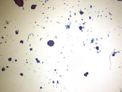
4x magnification of a 4R lymph node. Groups of cohesive epithelial appearing cells can be seen on low power. Lymphoid tissue is not easily identified.
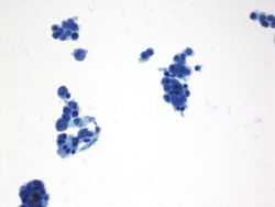
20x magnification of a 4R lymph node. This is a cellular specimen with groups of cells along what appear to be a papillary or papillary-like structure. Single cells are also dispersed in the background. The cells are haphazardly arranged.
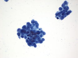
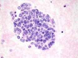
40x magnification of a 4R lymph node. On higher power, the nuclei appear mildly atypical and the cytoplasm is delicate and finely vacuolated. The nuclear contours are somewhat irregular.
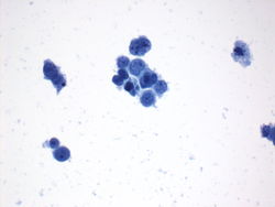
Cell block of 4R lymph node. The cytoplasm does not appear as vacuolated on alcohol fixed cell block material, but the nuclei are relatively uniform, but somewhat atypical.
Cell block of 4R lymph node. The cytoplasm does not appear as vacuolated on alcohol fixed cell block material, but the nuclei are relatively uniform, but somewhat atypical.


Immunohistochemistry
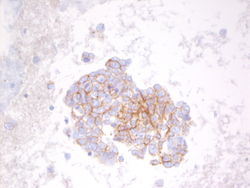
CD56 on pleural fluid shows positive cytoplasmic staining.
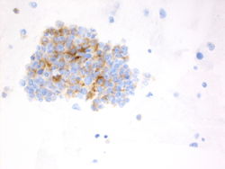
Synaptophysin on pleural fluid shows positive cytoplasmic staining.


Resident Questions
Final Diagnosis
Cytology
- Rapid diagnosis: Non-small cell carcinoma.
- Final diagnosis: Renal cell carcinoma.
Case Discussion
This is a classic case of metastatic renal cell carcinoma.
| ||||||||