Cytologically Yours: CoW: 20131209: Difference between revisions
Jump to navigation
Jump to search
No edit summary |
No edit summary |
||
| Line 1: | Line 1: | ||
== Clinical Summary == | == Clinical Summary == | ||
The patient is an | The patient is an 64 year old white male who presented with left sided back pain. Imaging showed a left perinephric retroperitoneal hematoma and a left renal lower pole cystic lesion with hemorrhage. Additional imaging showed numerous pulmonary lesions. A endobronchial ultrasound guided fine needle aspiration was scheduled. | ||
=== Past Medical History === | === Past Medical History === | ||
* | * Congestive heart failure | ||
* | * Ventricular tachycardia | ||
* | * Ischemic heart disease | ||
=== Past Surgical History === | === Past Surgical History === | ||
* | * Coronary stent placement | ||
* | * Implant of AICD | ||
===Clinical Plan=== | ===Clinical Plan=== | ||
The | The concern is a primary renal malignancy with metastatic disease to lungs. An endobronchial ultrasound guided FNA is scheduled. | ||
==Radiology== | ==Radiology== | ||
* | * CT Abdomen shows a large perinephric hematoma and large low anterior structure in left lower pole suspicious for a hemorrhagic renal cell carcinoma. | ||
* CT | * CT Chest shows multiple small lung lesions measuring up to 13x12 mm in greatest dimension. | ||
==Pathology== | ==Pathology== | ||
| Line 52: | Line 49: | ||
==Case Discussion== | ==Case Discussion== | ||
This is a classic case of | This is a classic case of metastatic renal cell carcinoma. | ||
</div></div> | </div></div> | ||
Revision as of 15:53, 14 January 2014
Clinical Summary
The patient is an 64 year old white male who presented with left sided back pain. Imaging showed a left perinephric retroperitoneal hematoma and a left renal lower pole cystic lesion with hemorrhage. Additional imaging showed numerous pulmonary lesions. A endobronchial ultrasound guided fine needle aspiration was scheduled.
Past Medical History
- Congestive heart failure
- Ventricular tachycardia
- Ischemic heart disease
Past Surgical History
- Coronary stent placement
- Implant of AICD
Clinical Plan
The concern is a primary renal malignancy with metastatic disease to lungs. An endobronchial ultrasound guided FNA is scheduled.
Radiology
- CT Abdomen shows a large perinephric hematoma and large low anterior structure in left lower pole suspicious for a hemorrhagic renal cell carcinoma.
- CT Chest shows multiple small lung lesions measuring up to 13x12 mm in greatest dimension.
Pathology
Cytology
-
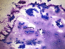 4x magnification of a 4R lymph node. There is a polymorphic lymphoid population with scattered large atypical cells.
 -
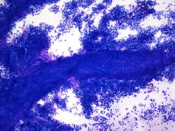 20x magnification of paracaval lymph node. There are small lymphocytes with background lymphoglandular bodies. Scattered eosinophils and large atypical cells with prominent nucleoli.
 -
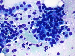 40x magnification of paracaval lymph node. There are atypical binucleated cells among the large atypical cells.
 -
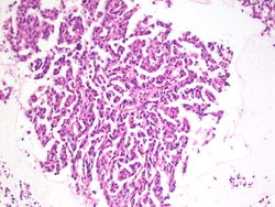 40x magnification of paracaval lymph node. There are atypical binucleated cells among the large atypical cells.
 -
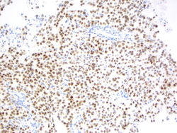 40x magnification of paracaval lymph node. There are atypical binucleated cells among the large atypical cells.


Resident Questions
Final Diagnosis
Cytology
- Positive for malignancy, the differential diagnosis includes melanoma and Hodgkin lymphoma.
Biopsy
- Classical Hodgkin lymphoma, favor mixed type.
Case Discussion
This is a classic case of metastatic renal cell carcinoma.
| ||||||||